• 文献检索
  • 文档翻译
  • 深度研究
  • 学术资讯
  • Suppr Zotero 插件Zotero 插件
  • 邀请有礼
  • 套餐&价格
  • 历史记录
应用&插件
Suppr Zotero 插件Zotero 插件浏览器插件Mac 客户端Windows 客户端微信小程序
定价
高级版会员购买积分包购买API积分包
服务
文献检索文档翻译深度研究API 文档MCP 服务
关于我们
关于 Suppr公司介绍联系我们用户协议隐私条款
关注我们

Suppr 超能文献

核心技术专利:CN118964589B侵权必究
粤ICP备2023148730 号-1Suppr @ 2026

文献检索

告别复杂PubMed语法,用中文像聊天一样搜索,搜遍4000万医学文献。AI智能推荐,让科研检索更轻松。

立即免费搜索

文件翻译

保留排版,准确专业,支持PDF/Word/PPT等文件格式,支持 12+语言互译。

免费翻译文档

深度研究

AI帮你快速写综述,25分钟生成高质量综述,智能提取关键信息,辅助科研写作。

立即免费体验

各种非增强磁共振血管造影(MRA)技术对配备Surpass Evolve血流分流器的血管流动模型中管腔可视性的影响。

Impact of Various Non-Contrast-Enhanced MRA Techniques on Lumen Visibility in Vascular Flow Models with a Surpass Evolve Flow Diverter.

作者信息

Ozpeynirci Yigit, Gorodezky Margarita, Sanches Augusto Fava, Mandava Sagar, Solana Ana Beatriz, Liebig Thomas

机构信息

Institute for Diagnostic and Interventional Neuroradiology, University Hospital, Ludwig-Maximilians-University (LMU), Marchioninistr. 15, 81377 Munich, Germany.

GE HealthCare, 80807 Munich, Germany.

出版信息

Diagnostics (Basel). 2024 May 30;14(11):1146. doi: 10.3390/diagnostics14111146.

DOI:10.3390/diagnostics14111146
PMID:38893675
原文链接:https://pmc.ncbi.nlm.nih.gov/articles/PMC11171646/
Abstract

BACKGROUND

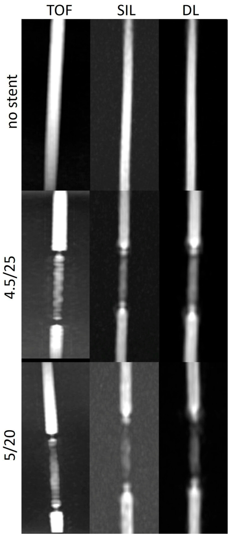
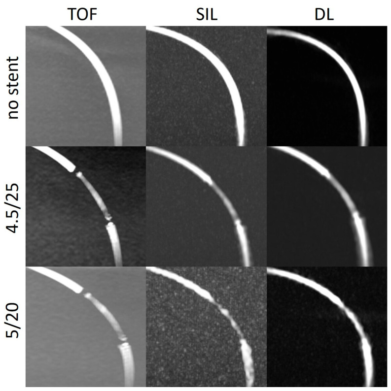
Silent MRA has shown promising results in evaluating the stents used for intracranial aneurysm treatment. A deep learning-based denoising and deranging algorithm was recently introduced by GE HealthCare. The purpose of this study was to compare the performance of several MRA techniques regarding lumen visibility in silicone models with flow diverter stents.

METHODS

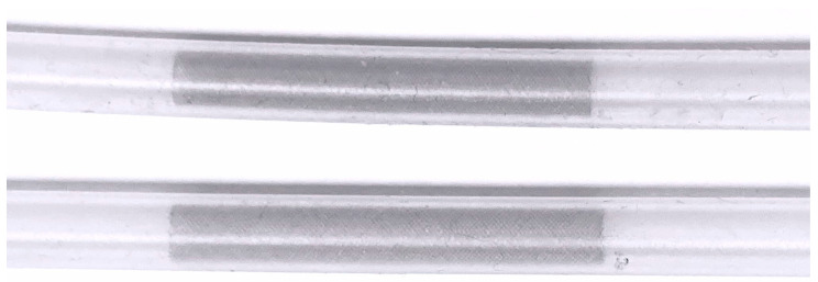
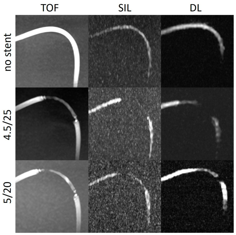
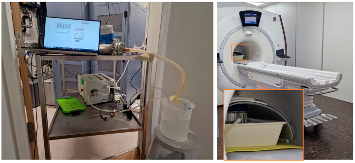
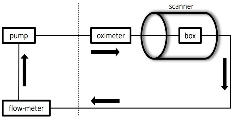
Two Surpass Evolve stents of different sizes were implanted in two silicone tubes. The tubes were placed in separate boxes in the straight position and in two different curve configurations and connected to a pulsatile pump to construct a flow loop. Using a 3.0T MRI scanner, TOF and silent MRA images were acquired, and deep learning reconstruction was applied to the silent MRA dataset. The intraluminal signal intensity in the stent (SI), in the tube outside the stent (SI), and of the background (SI) were measured for each scan.

RESULTS

The SI/SI and SI/SI ratios were higher in the silent scans and DL-based reconstructions than in the TOF images. The stent tips created severe artefacts in the TOF images, which could not be observed in the silent scans.

CONCLUSIONS

Our study demonstrates that the DL reconstruction algorithm improves the quality of the silent MRA technique in evaluating the flow diverter stent patency.

摘要

背景

静态磁共振血管造影(MRA)在评估用于颅内动脉瘤治疗的支架方面已显示出有前景的结果。通用电气医疗集团最近推出了一种基于深度学习的去噪和去伪影算法。本研究的目的是比较几种MRA技术在带有血流导向支架的硅胶模型中关于管腔可视性的性能。

方法

将两个不同尺寸的Surpass Evolve支架植入两根硅胶管中。将管子笔直放置以及以两种不同的弯曲构型分别置于单独的盒子中,并连接到脉动泵以构建血流环路。使用3.0T磁共振成像(MRI)扫描仪采集时间飞跃法(TOF)和静态MRA图像,并将深度学习重建应用于静态MRA数据集。对每次扫描测量支架内的腔内信号强度(SI)、支架外管内的信号强度(SI)以及背景的信号强度(SI)。

结果

与TOF图像相比,静态扫描和基于深度学习的重建中的SI/SI和SI/SI比值更高。支架末端在TOF图像中产生严重伪影,而在静态扫描中未观察到。

结论

我们的研究表明,深度学习重建算法在评估血流导向支架通畅性方面提高了静态MRA技术的质量。

https://cdn.ncbi.nlm.nih.gov/pmc/blobs/cb79/11171646/f3f7d3491df3/diagnostics-14-01146-g006.jpg
https://cdn.ncbi.nlm.nih.gov/pmc/blobs/cb79/11171646/a50c059e7d9d/diagnostics-14-01146-g001.jpg
https://cdn.ncbi.nlm.nih.gov/pmc/blobs/cb79/11171646/fb4fe98ac861/diagnostics-14-01146-g002.jpg
https://cdn.ncbi.nlm.nih.gov/pmc/blobs/cb79/11171646/c1b85de87895/diagnostics-14-01146-g003.jpg
https://cdn.ncbi.nlm.nih.gov/pmc/blobs/cb79/11171646/f9b8b704151f/diagnostics-14-01146-g004.jpg
https://cdn.ncbi.nlm.nih.gov/pmc/blobs/cb79/11171646/45c38b0d0655/diagnostics-14-01146-g005.jpg
https://cdn.ncbi.nlm.nih.gov/pmc/blobs/cb79/11171646/f3f7d3491df3/diagnostics-14-01146-g006.jpg

相似文献

1
Impact of Various Non-Contrast-Enhanced MRA Techniques on Lumen Visibility in Vascular Flow Models with a Surpass Evolve Flow Diverter.各种非增强磁共振血管造影(MRA)技术对配备Surpass Evolve血流分流器的血管流动模型中管腔可视性的影响。
Diagnostics (Basel). 2024 May 30;14(11):1146. doi: 10.3390/diagnostics14111146.
2
Assessing Blood Flow in an Intracranial Stent: A Feasibility Study of MR Angiography Using a Silent Scan after Stent-Assisted Coil Embolization for Anterior Circulation Aneurysms.评估颅内支架内的血流:一项关于支架辅助弹簧圈栓塞治疗前循环动脉瘤后使用静音扫描进行磁共振血管造影的可行性研究。
AJNR Am J Neuroradiol. 2015 May;36(5):967-70. doi: 10.3174/ajnr.A4199. Epub 2014 Dec 18.
3
Predicting the O'Kelly-Marotta scale score after flow-diverter stent placement using silent MRA.使用静息 MRA 预测血流导向装置置入术后 O'Kelly-Marotta 评分。
Jpn J Radiol. 2024 Dec;42(12):1403-1412. doi: 10.1007/s11604-024-01632-1. Epub 2024 Aug 29.
4
Usefulness of Non-Contrast-Enhanced MR Angiography Using a Silent Scan for Follow-Up after Y-Configuration Stent-Assisted Coil Embolization for Basilar Tip Aneurysms.使用静音扫描的非增强磁共振血管造影在Y形支架辅助弹簧圈栓塞基底动脉尖动脉瘤后随访中的应用价值
AJNR Am J Neuroradiol. 2017 Mar;38(3):577-581. doi: 10.3174/ajnr.A5033. Epub 2016 Dec 22.
5
Non-Contrast-Enhanced Silent Scan MR Angiography of Intracranial Anterior Circulation Aneurysms Treated with a Low-Profile Visualized Intraluminal Support Device.使用低轮廓可视化腔内支撑装置治疗的颅内前循环动脉瘤的非增强磁共振血管造影静息扫描
AJNR Am J Neuroradiol. 2017 Aug;38(8):1610-1616. doi: 10.3174/ajnr.A5223. Epub 2017 May 18.
6
Magnetic Resonance Angiography After Flow Diversion: The Use of Complementary Magnetic Resonance Angiography Techniques to Monitor Aneurysm Occlusion and Device and Arterial Branch Patency After Flow Diverter Placement.血流导向装置治疗后磁共振血管成像:应用互补性磁共振血管成像技术监测血流导向装置置入后动脉瘤闭塞和装置及载瘤动脉分支通畅情况。
World Neurosurg. 2022 Jun;162:e147-e155. doi: 10.1016/j.wneu.2022.02.096. Epub 2022 Mar 3.
7
[Visualization of Four Intracranial Stents and In-stent Stenosis with Stenotic Models by 3D-TOF-MRA].[通过三维时间飞跃法磁共振血管造影术对四个颅内支架及伴有狭窄模型的支架内狭窄进行可视化研究]
Nihon Hoshasen Gijutsu Gakkai Zasshi. 2019;75(8):747-754. doi: 10.6009/jjrt.2019_JSRT_75.8.747.
8
Usefulness of Silent MRA for Evaluation of Aneurysm after Stent-Assisted Coil Embolization.支架辅助线圈栓塞后评估动脉瘤时使用 Silent MRA 的实用性。
Korean J Radiol. 2022 Feb;23(2):246-255. doi: 10.3348/kjr.2021.0332. Epub 2022 Jan 4.
9
Usefulness of Silent MR Angiography for Intracranial Aneurysms Treated with a Flow-Diverter Device.密网支架治疗颅内动脉瘤后应用静默磁共振血管成像的价值
AJNR Am J Neuroradiol. 2019 May;40(5):808-814. doi: 10.3174/ajnr.A6047. Epub 2019 May 2.
10
Placement of a Stent within a Flow Diverter Improves Aneurysm Occlusion Rates.支架在血流导向装置内的放置可提高动脉瘤闭塞率。
AJNR Am J Neuroradiol. 2019 Nov;40(11):1932-1938. doi: 10.3174/ajnr.A6237. Epub 2019 Oct 3.

本文引用的文献

1
Usefulness of Silent MRA for Evaluation of Aneurysm after Stent-Assisted Coil Embolization.支架辅助线圈栓塞后评估动脉瘤时使用 Silent MRA 的实用性。
Korean J Radiol. 2022 Feb;23(2):246-255. doi: 10.3348/kjr.2021.0332. Epub 2022 Jan 4.
2
Investigating Signal Loss due to a Carotid Artery Stent in 3D-TOF-MRA.探讨 3D-TOF-MRA 中颈动脉支架导致的信号丢失。
Magn Reson Med Sci. 2021 Sep 1;20(3):303-311. doi: 10.2463/mrms.mp.2019-0083. Epub 2020 Sep 18.
3
Silent MRI: A Future of Less Intrusive and More Diagnostic Scanning.
Acad Radiol. 2020 Apr;27(4):550-551. doi: 10.1016/j.acra.2020.01.032. Epub 2020 Feb 24.
4
Usefulness of Silent MR Angiography for Intracranial Aneurysms Treated with a Flow-Diverter Device.密网支架治疗颅内动脉瘤后应用静默磁共振血管成像的价值
AJNR Am J Neuroradiol. 2019 May;40(5):808-814. doi: 10.3174/ajnr.A6047. Epub 2019 May 2.
5
Neurovascular stent artifacts in 3D-TOF and 3D-PCMRI: Influence of stent design on flow measurement.三维时间飞跃法和三维对比增强磁共振血管成像中的神经血管支架伪影:支架设计对血流测量的影响。
Magn Reson Med. 2019 Jan;81(1):560-572. doi: 10.1002/mrm.27352. Epub 2018 Jun 12.
6
Clinical Evaluation of Silent T1-Weighted MRI and Silent MR Angiography of the Brain.脑的静息 T1 加权 MRI 和静息 MR 血管成像的临床评估。
AJR Am J Roentgenol. 2018 Feb;210(2):404-411. doi: 10.2214/AJR.17.18247. Epub 2017 Nov 7.
7
Non-Contrast-Enhanced Silent Scan MR Angiography of Intracranial Anterior Circulation Aneurysms Treated with a Low-Profile Visualized Intraluminal Support Device.使用低轮廓可视化腔内支撑装置治疗的颅内前循环动脉瘤的非增强磁共振血管造影静息扫描
AJNR Am J Neuroradiol. 2017 Aug;38(8):1610-1616. doi: 10.3174/ajnr.A5223. Epub 2017 May 18.
8
Usefulness of Non-Contrast-Enhanced MR Angiography Using a Silent Scan for Follow-Up after Y-Configuration Stent-Assisted Coil Embolization for Basilar Tip Aneurysms.使用静音扫描的非增强磁共振血管造影在Y形支架辅助弹簧圈栓塞基底动脉尖动脉瘤后随访中的应用价值
AJNR Am J Neuroradiol. 2017 Mar;38(3):577-581. doi: 10.3174/ajnr.A5033. Epub 2016 Dec 22.
9
Assessing Blood Flow in an Intracranial Stent: A Feasibility Study of MR Angiography Using a Silent Scan after Stent-Assisted Coil Embolization for Anterior Circulation Aneurysms.评估颅内支架内的血流:一项关于支架辅助弹簧圈栓塞治疗前循环动脉瘤后使用静音扫描进行磁共振血管造影的可行性研究。
AJNR Am J Neuroradiol. 2015 May;36(5):967-70. doi: 10.3174/ajnr.A4199. Epub 2014 Dec 18.
10
Follow-up of intracranial aneurysms treated by flow diverter: comparison of three-dimensional time-of-flight MR angiography (3D-TOF-MRA) and contrast-enhanced MR angiography (CE-MRA) sequences with digital subtraction angiography as the gold standard.血流导向装置治疗颅内动脉瘤的随访:以数字减影血管造影为金标准比较三维时间飞跃磁共振血管造影(3D-TOF-MRA)和对比增强磁共振血管造影(CE-MRA)序列
J Neurointerv Surg. 2016 Jan;8(1):81-6. doi: 10.1136/neurintsurg-2014-011449. Epub 2014 Oct 28.