Xu Minghe, Li Bo, Wang Shuang, Chen Chunlian, Liu Zhe, Ji Yuqing, Liu Kai, Niu Yujun
Jinzhou Medical University, Jinzhou, China.
Department of Radiology, The 960th Hospital of People's Liberation Army Joint Logistic Support Force, Jinan, China.
Front Psychiatry. 2024 Jun 4;15:1364713. doi: 10.3389/fpsyt.2024.1364713. eCollection 2024.
Chronic insomnia disorder (CID) is usually associated with Generalized Anxiety Disorder (GAD), which may change brain structure and function. However, the possible brain markers, imaging characteristics, and pathophysiology are unknown.
To look at the probable brain markers, imaging characteristics, and pathogenesis of CID in combination with GAD.
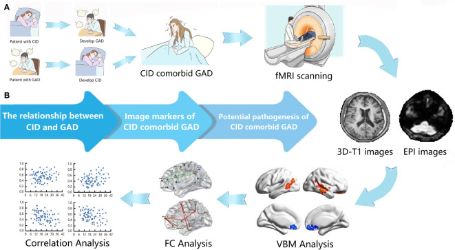
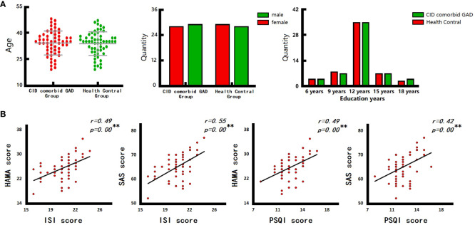
A total of 57 patients with CID concomitant GAD and 57 healthy controls (HC) were enrolled. Voxel-based morphometry (VBM) and functional connectivity (FC) were utilized to measure gray matter volume (GMV) and functional changes. Correlation analysis was utilized to identify relationships between brain changes and clinical characteristics.
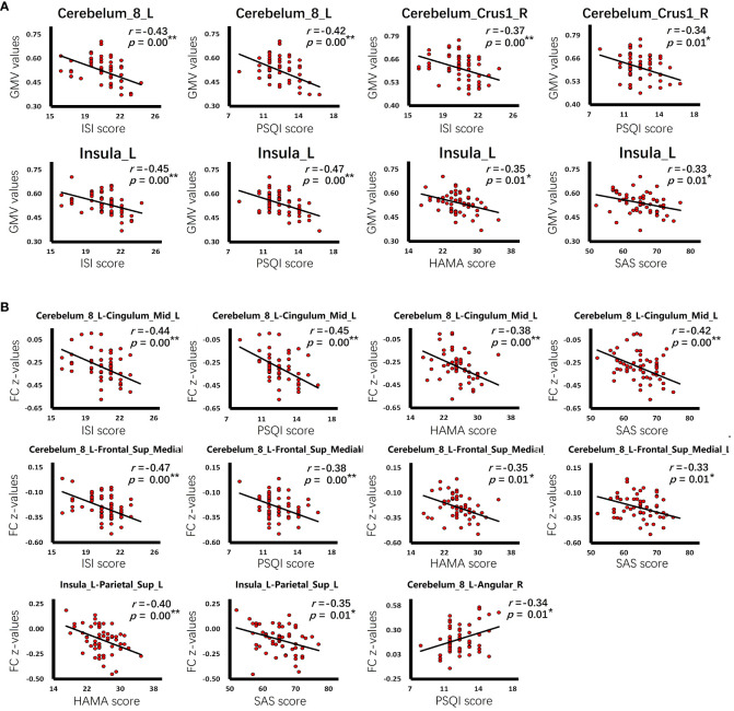
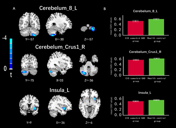
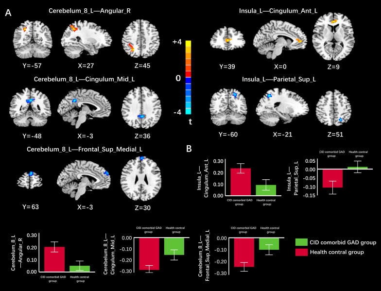
Patients had decreased GMV in the left cerebellum, right cerebellar peduncle, and left insula; increased FC between the left cerebellum and right angular gyrus, as well as between the left insula and anterior left cingulate gyrus; and decreased FC in several areas, including the left cerebellum with the middle left cingulate gyrus and the left insula with the left superior postcentral gyrus. These brain changes related to CID and GAD. These data could be used to identify relevant brain markers, imaging features, and to better understand the etiology.
The intensity of insomnia in patients was strongly related to the severity of anxiety. The lower GMV in the cerebellum could be interpreted as an imaging characteristic of CID. Reduced GMV in the insula, as well as aberrant function in the cingulate gyrus and prefrontal lobe, may contribute to the pathophysiology of CID and GAD. Abnormal function in the postcentral gyrus and angular gyrus may be associated with patients' clinical complaints.
慢性失眠障碍(CID)通常与广泛性焦虑障碍(GAD)相关,这可能会改变大脑结构和功能。然而,潜在的脑标志物、影像学特征和病理生理学尚不清楚。
探讨合并GAD的CID可能的脑标志物、影像学特征及发病机制。
共纳入57例合并GAD的CID患者和57名健康对照(HC)。采用基于体素的形态学测量(VBM)和功能连接(FC)来测量灰质体积(GMV)和功能变化。利用相关性分析来确定脑变化与临床特征之间的关系。
患者左侧小脑、右侧小脑脚和左侧岛叶的GMV降低;左侧小脑与右侧角回之间以及左侧岛叶与左侧前扣带回之间的FC增加;包括左侧小脑与左侧中部扣带回以及左侧岛叶与左侧中央后回上部之间在内的几个区域的FC降低。这些脑变化与CID和GAD相关。这些数据可用于识别相关脑标志物、影像学特征,并更好地理解病因。
患者失眠的严重程度与焦虑的严重程度密切相关。小脑GMV降低可被解释为CID的影像学特征。岛叶GMV降低以及扣带回和前额叶功能异常可能导致CID和GAD 的病理生理过程。中央后回和角回的功能异常可能与患者的临床症状有关。