Park Jang Woo, Tian Yunan, Kim Sang-Tae, Park Chanwoo, Kim Yu Mi, Chung Hye Kyung, Kim Kyeong Min, Jahng Geon-Ho
Korea Radioisotope Center for Pharmaceuticals, Korea Institute of Radiological and Medical Sciences, Seoul, Republic of Korea.
Department of Medicine, Graduate School, Kyung Hee University College of Medicine, Seoul, Republic of Korea.
Front Pharmacol. 2024 Jun 4;15:1392729. doi: 10.3389/fphar.2024.1392729. eCollection 2024.
Oligomeric amyloid beta (oAβ) is a toxic factor that acts in the early stage of Alzheimer's disease (AD) and may initiate the pathologic cascade. Therefore, detecting oAβ has a crucial role in the early diagnosis, monitoring, and treatment of AD.
The purpose of this study was to evaluate MRI signal changes in different mouse models and the time-dependent signal changes using our novel gadolinium (Gd)-dodecane tetraacetic acid (DOTA)- ob5 aptamer contrast agent.
We developed an MRI contrast agent by conjugating Gd-DOTA-DNA aptamer called ob5 to evaluate its ability to detect oAβ deposits in the brain using MRI. A total of 10 control mice, 9 3xTg AD mice, and 11 APP/PS/Tau AD mice were included in this study, with the age of each model being 16 or 36 weeks. A T1-weighted image was acquired at the time points before (0 min) and after injection of the contrast agent at 5, 10, 15, 20, and 25 min. The analyses were performed to compare MRI signal differences among the three groups and the time-dependent signal differences in different mouse models.
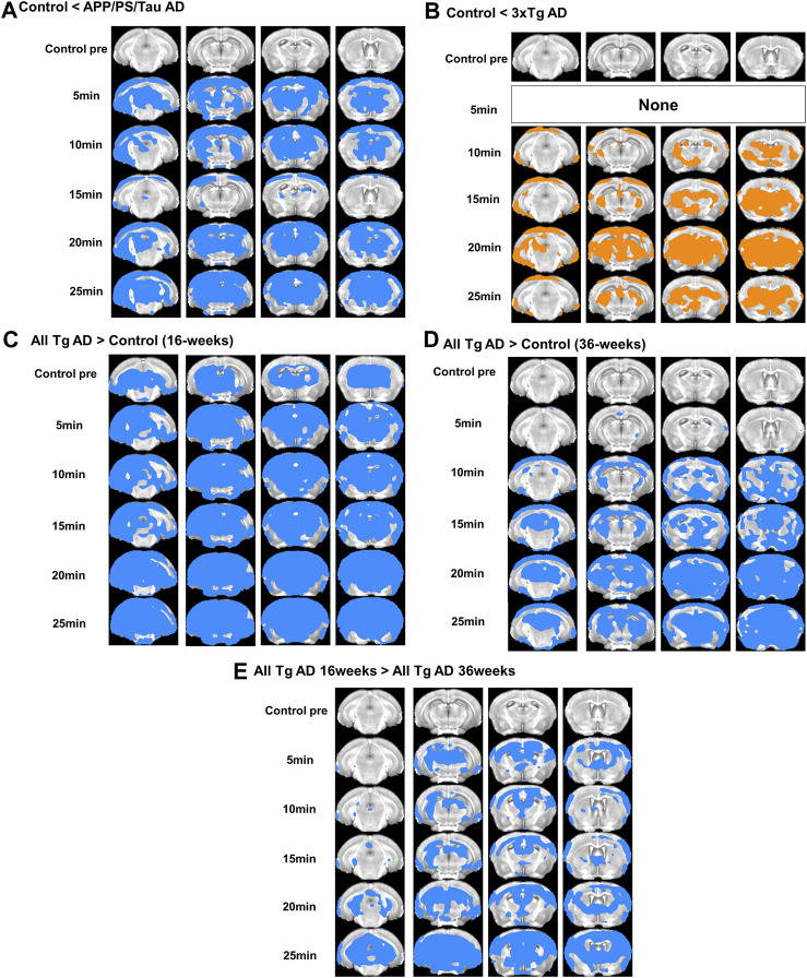
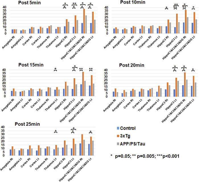
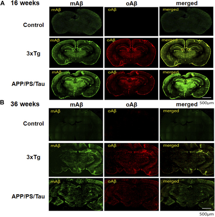
Both 3xTg AD and APP/PS/Tau AD mouse models had higher signal enhancement than control mice at all scan-time points after injection of our contrast media, especially in bilateral hippocampal areas. In particular, all Tg AD mouse models aged 16 weeks showed a higher contrast enhancement than those aged 36 weeks. For 3xTg AD and APP/PS/Tau AD groups, the signal enhancement was significantly different among the five time points (0 min, 5 min, 10 min, 15 min, 20 min, and 25 min) in multiple ROI areas, typically in the bilateral hippocampus, left thalamus, and left amygdala.
The findings of this study suggest that the expression of the contrast agent in different AD models demonstrates its translational flexibility across different species. The signal enhancement peaked around 15-20 min after injection of the contrast agent. Therefore, our novel contrast agent targeting oAβ has the potential ability to diagnose early AD and monitor the progression of AD.
寡聚淀粉样β蛋白(oAβ)是一种在阿尔茨海默病(AD)早期起作用的毒性因子,可能引发病理级联反应。因此,检测oAβ在AD的早期诊断、监测和治疗中具有关键作用。
本研究的目的是使用我们新型的钆(Gd)-十二烷四乙酸(DOTA)-ob5适配体造影剂,评估不同小鼠模型中的MRI信号变化以及随时间变化的信号变化。
我们通过将名为ob5的Gd-DOTA-DNA适配体偶联来开发一种MRI造影剂,以评估其使用MRI检测脑内oAβ沉积物的能力。本研究共纳入10只对照小鼠、9只3xTg AD小鼠和11只APP/PS/Tau AD小鼠,每个模型的年龄为16周或36周。在注射造影剂前(0分钟)以及注射后5、10、15、20和25分钟的时间点采集T1加权图像。进行分析以比较三组之间的MRI信号差异以及不同小鼠模型中随时间变化的信号差异。
在注射我们的造影剂后的所有扫描时间点,3xTg AD和APP/PS/Tau AD小鼠模型的信号增强均高于对照小鼠,尤其是在双侧海马区域。特别是,所有16周龄的Tg AD小鼠模型的对比增强均高于36周龄的模型。对于3xTg AD和APP/PS/Tau AD组,在多个感兴趣区(ROI),通常是双侧海马、左侧丘脑和左侧杏仁核,五个时间点(0分钟、5分钟、10分钟、15分钟、20分钟和25分钟)之间的信号增强存在显著差异。
本研究结果表明,造影剂在不同AD模型中的表达证明了其在不同物种间的转化灵活性。注射造影剂后约15 - 20分钟信号增强达到峰值。因此,我们新型的靶向oAβ的造影剂具有早期诊断AD和监测AD进展的潜在能力。