Dubowitz L M, Bydder G M, Mushin J
Arch Dis Child. 1985 Apr;60(4):349-55. doi: 10.1136/adc.60.4.349.
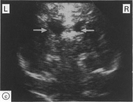
The evolution of severe periventricular leukomalacia was followed by ultrasonography in three newborn infants, and the subsequent myelination of the brain was assessed by nuclear magnetic resonance imaging. Four stages of periventricular leukomalacia could be identified by ultrasonography; (1) initial congestion, followed by (2) relative normalisation, (3) development of cysts, and (4) resolution of cysts but development of ventricular enlargement. All infants exhibited abnormal neurological signs from 36 weeks conceptual age and had unequivocal signs of cerebral palsy by 6 to 9 months of age. One infant became cortically blind but the other two seemed to have normal vision. Nuclear magnetic resonance imaging showed some abnormality of the ventricular system and delayed myelination in all three infants. The delay was most noticeable in the opticothalamic region, which was also the site of the most extensive lesions observed on ultrasonography. Progress in myelination was observed in the infants where a repeat scan was performed.
对3例新生儿进行超声检查以追踪重度脑室周围白质软化的演变过程,并通过核磁共振成像评估随后的脑髓鞘形成情况。超声检查可识别出脑室周围白质软化的四个阶段:(1)最初的充血,随后是(2)相对正常化,(3)囊肿形成,以及(4)囊肿消退但脑室扩大。所有婴儿在孕36周时均出现异常神经体征,在6至9个月大时均有明确的脑瘫体征。一名婴儿出现皮质盲,但另外两名婴儿视力似乎正常。核磁共振成像显示,所有三名婴儿的脑室系统均有一些异常,且髓鞘形成延迟。这种延迟在视丘区最为明显,而视丘区也是超声检查中观察到病变最广泛的部位。对进行重复扫描的婴儿观察到了髓鞘形成的进展。