Steiner Dylan, Sultan Lila, Sullivan Travis, Liu Hanqiao, Zhang Sherry, LeClerc Ashley, Alekseyev Yuriy O, Liu Gang, Mazzilli Sarah A, Zhang Jiarui, Rieger-Christ Kimberly, Burks Eric J, Beane Jennifer, Lenburg Marc E
Department of Medicine, Section of Computational Biomedicine, Boston University Chobanian and Avedisian School of Medicine, Boston, MA, USA.
Department of Pathology and Laboratory Medicine, Boston University Chobanian and Avedisian School of Medicine, Boston, MA, USA.
bioRxiv. 2024 Jun 10:2024.06.07.597993. doi: 10.1101/2024.06.07.597993.
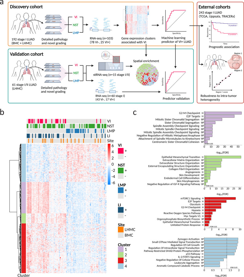
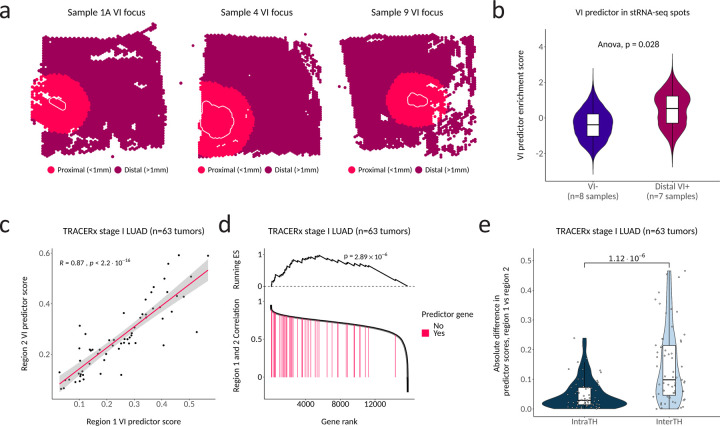
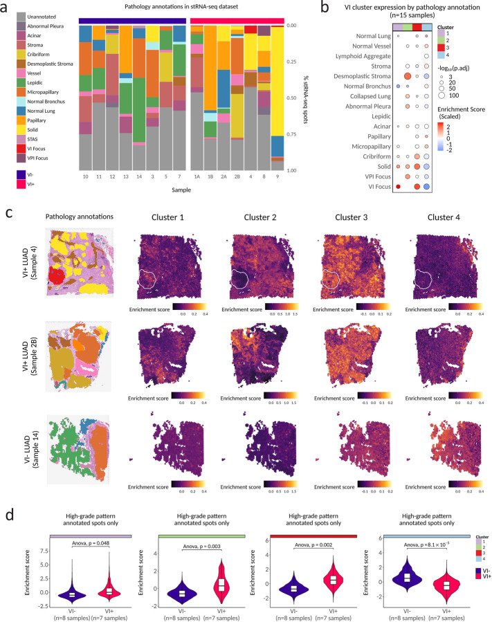
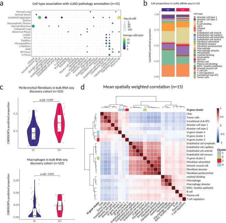
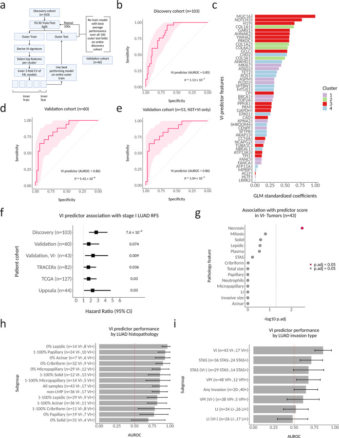
Microscopic vascular invasion (VI) is predictive of recurrence and benefit from lobectomy in stage I lung adenocarcinoma (LUAD) but is difficult to assess in resection specimens and cannot be accurately predicted prior to surgery. Thus, new biomarkers are needed to identify this aggressive subset of stage I LUAD tumors. To assess molecular and microenvironment features associated with angioinvasive LUAD we profiled 162 resected stage I tumors with and without VI by RNA-seq and explored spatial patterns of gene expression in a subset of 15 samples by high-resolution spatial transcriptomics (stRNA-seq). Despite the small size of invaded blood vessels, we identified a gene expression signature of VI from the bulk RNA-seq discovery cohort (n=103) and found that it was associated with VI foci, desmoplastic stroma, and high-grade patterns in our stRNA-seq data. We observed a stronger association with high-grade patterns from VI compared with VI tumors. Using the discovery cohort, we developed a transcriptomic predictor of VI, that in an independent validation cohort (n=60) was associated with VI (AUROC=0.86; p=5.42×10) and predictive of recurrence-free survival (HR=1.98; p=0.024), even in VI LUAD (HR=2.76; p=0.003). To determine our VI predictor's robustness to intra-tumor heterogeneity we used RNA-seq data from multi-region sampling of stage I LUAD cases in TRACERx, where the predictor scores showed high correlation (R=0.87, p<2.2×10) between two randomly sampled regions of the same tumor. Our study suggests that VI-associated gene expression changes are detectable beyond the site of intravasation and can be used to predict the presence of VI. This may enable the prediction of angioinvasive LUAD from biopsy specimens, allowing for more tailored medical and surgical management of stage I LUAD.
微脉管浸润(VI)可预测I期肺腺癌(LUAD)的复发及肺叶切除的获益情况,但在切除标本中难以评估,且术前无法准确预测。因此,需要新的生物标志物来识别这一侵袭性较强的I期LUAD肿瘤亚组。为评估与血管侵袭性LUAD相关的分子和微环境特征,我们通过RNA测序对162例有或无VI的I期切除肿瘤进行了分析,并通过高分辨率空间转录组学(stRNA-seq)探索了15个样本亚组中的基因表达空间模式。尽管侵袭血管的尺寸较小,但我们从批量RNA测序发现队列(n = 103)中确定了VI的基因表达特征,并发现其与我们的stRNA-seq数据中的VI病灶、促结缔组织增生性基质和高级别模式相关。与无VI的肿瘤相比,我们观察到VI与高级别模式之间的关联更强。利用发现队列,我们开发了一种VI的转录组预测指标,在一个独立验证队列(n = 60)中,该指标与VI相关(曲线下面积=0.86;p = 5.42×10),并可预测无复发生存期(风险比=1.98;p = 0.024),即使在有VI的LUAD中也是如此(风险比=2.76;p = 0.003)。为确定我们的VI预测指标对肿瘤内异质性的稳健性,我们使用了TRACERx中I期LUAD病例多区域采样的RNA测序数据,在同一肿瘤的两个随机采样区域之间,预测指标得分显示出高度相关性(R = 0.87,p < 2.2×10)。我们的研究表明,VI相关的基因表达变化在血管内侵入部位之外即可检测到,并可用于预测VI的存在。这可能有助于从活检标本中预测血管侵袭性LUAD,从而为I期LUAD提供更具针对性的医疗和手术管理。