• 文献检索
  • 文档翻译
  • 深度研究
  • 学术资讯
  • Suppr Zotero 插件Zotero 插件
  • 邀请有礼
  • 套餐&价格
  • 历史记录
应用&插件
Suppr Zotero 插件Zotero 插件浏览器插件Mac 客户端Windows 客户端微信小程序
定价
高级版会员购买积分包购买API积分包
服务
文献检索文档翻译深度研究API 文档MCP 服务
关于我们
关于 Suppr公司介绍联系我们用户协议隐私条款
关注我们

Suppr 超能文献

核心技术专利:CN118964589B侵权必究
粤ICP备2023148730 号-1Suppr @ 2026

文献检索

告别复杂PubMed语法,用中文像聊天一样搜索,搜遍4000万医学文献。AI智能推荐,让科研检索更轻松。

立即免费搜索

文件翻译

保留排版,准确专业,支持PDF/Word/PPT等文件格式,支持 12+语言互译。

免费翻译文档

深度研究

AI帮你快速写综述,25分钟生成高质量综述,智能提取关键信息,辅助科研写作。

立即免费体验

利用 CytoSPACE 实现单细胞和空间转录组的高分辨率比对。

High-resolution alignment of single-cell and spatial transcriptomes with CytoSPACE.

机构信息

Institute for Stem Cell Biology and Regenerative Medicine, Stanford University, Stanford, CA, USA.

Department of Biomedical Data Science, Stanford University, Stanford, CA, USA.

出版信息

Nat Biotechnol. 2023 Nov;41(11):1543-1548. doi: 10.1038/s41587-023-01697-9. Epub 2023 Mar 6.

DOI:10.1038/s41587-023-01697-9
PMID:36879008
原文链接:https://pmc.ncbi.nlm.nih.gov/articles/PMC10635828/
Abstract

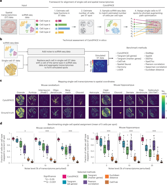
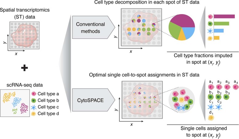
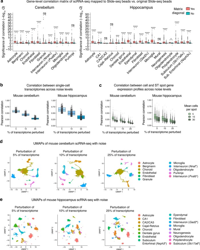
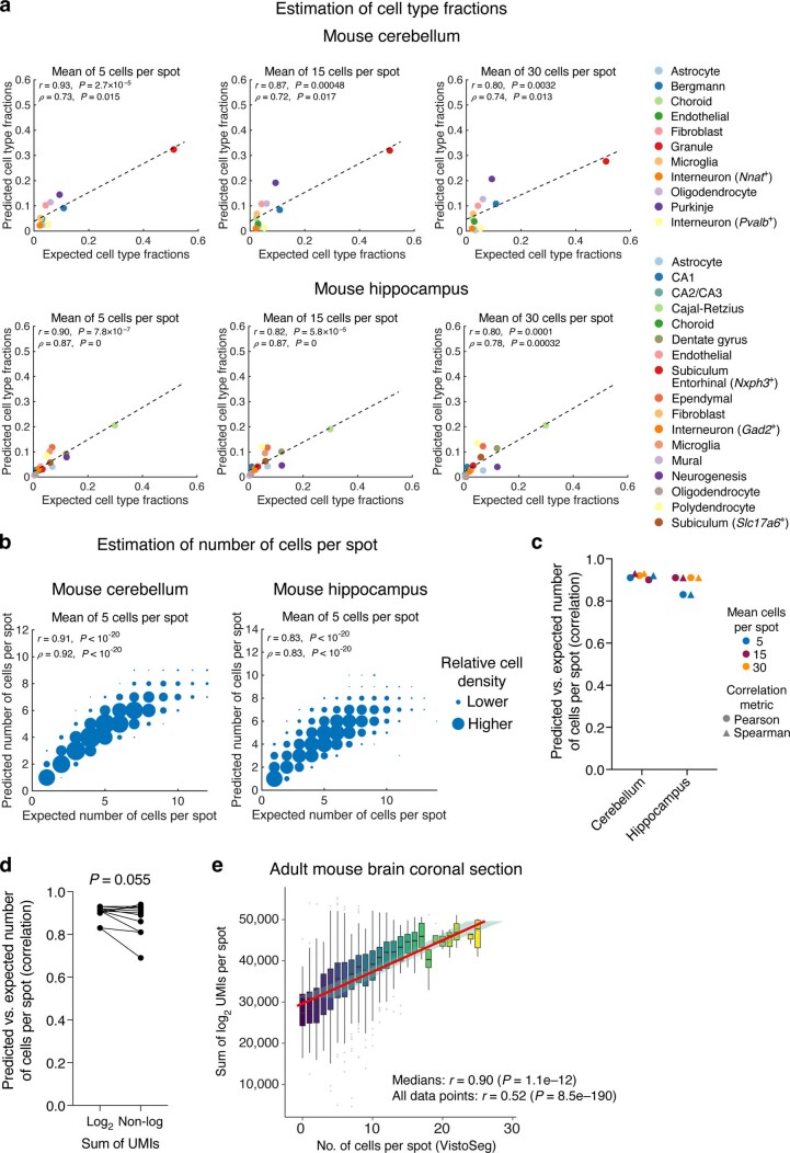
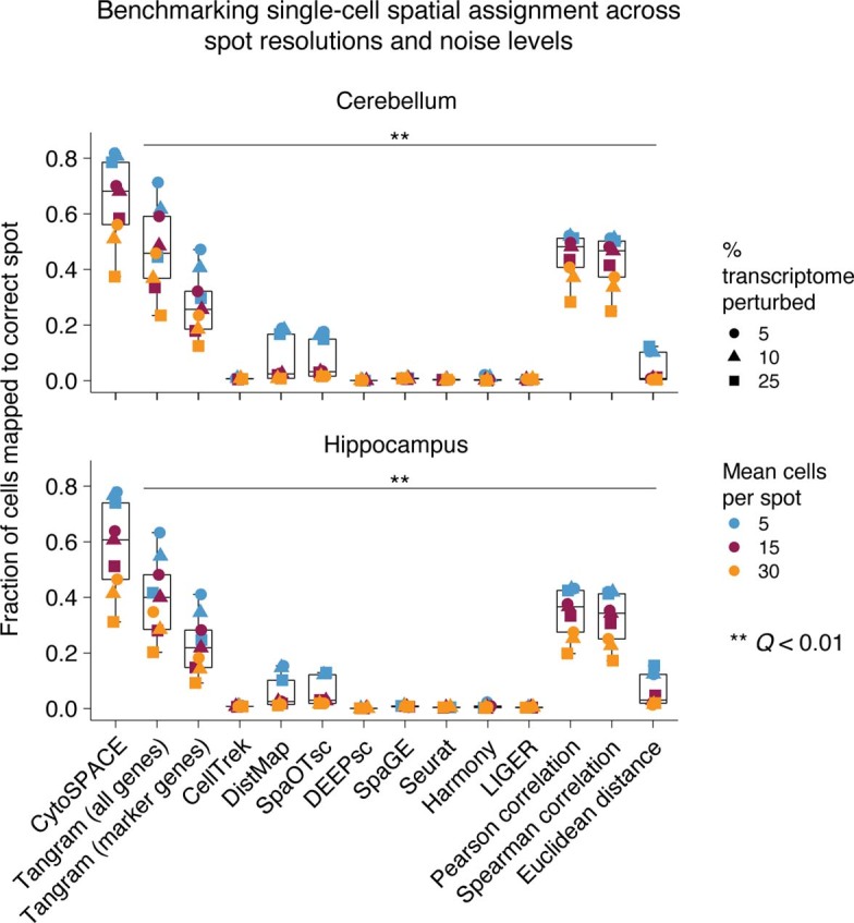
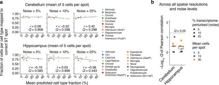
Recent studies have emphasized the importance of single-cell spatial biology, yet available assays for spatial transcriptomics have limited gene recovery or low spatial resolution. Here we introduce CytoSPACE, an optimization method for mapping individual cells from a single-cell RNA sequencing atlas to spatial expression profiles. Across diverse platforms and tissue types, we show that CytoSPACE outperforms previous methods with respect to noise tolerance and accuracy, enabling tissue cartography at single-cell resolution.

摘要

最近的研究强调了单细胞空间生物学的重要性,但现有的空间转录组学检测方法存在基因回收率有限或空间分辨率低的问题。在这里,我们介绍了 CytoSPACE,这是一种将单细胞 RNA 测序图谱中的单个细胞映射到空间表达图谱的优化方法。在不同的平台和组织类型中,我们表明 CytoSPACE 在噪声容忍度和准确性方面优于以前的方法,从而能够以单细胞分辨率进行组织绘图。

https://cdn.ncbi.nlm.nih.gov/pmc/blobs/0dfb/10635828/6cd7de3a32bc/41587_2023_1697_Fig12_ESM.jpg
https://cdn.ncbi.nlm.nih.gov/pmc/blobs/0dfb/10635828/da0c79c5f53d/41587_2023_1697_Fig1_HTML.jpg
https://cdn.ncbi.nlm.nih.gov/pmc/blobs/0dfb/10635828/5e79bcedcb16/41587_2023_1697_Fig2_HTML.jpg
https://cdn.ncbi.nlm.nih.gov/pmc/blobs/0dfb/10635828/5b76ec354745/41587_2023_1697_Fig3_ESM.jpg
https://cdn.ncbi.nlm.nih.gov/pmc/blobs/0dfb/10635828/1e75fbc92d6c/41587_2023_1697_Fig4_ESM.jpg
https://cdn.ncbi.nlm.nih.gov/pmc/blobs/0dfb/10635828/5ec56d86302c/41587_2023_1697_Fig5_ESM.jpg
https://cdn.ncbi.nlm.nih.gov/pmc/blobs/0dfb/10635828/8b3f66369941/41587_2023_1697_Fig6_ESM.jpg
https://cdn.ncbi.nlm.nih.gov/pmc/blobs/0dfb/10635828/4ae5cd6a4a76/41587_2023_1697_Fig7_ESM.jpg
https://cdn.ncbi.nlm.nih.gov/pmc/blobs/0dfb/10635828/a7af71e88a10/41587_2023_1697_Fig8_ESM.jpg
https://cdn.ncbi.nlm.nih.gov/pmc/blobs/0dfb/10635828/e6dc644381be/41587_2023_1697_Fig9_ESM.jpg
https://cdn.ncbi.nlm.nih.gov/pmc/blobs/0dfb/10635828/090f9f76f102/41587_2023_1697_Fig10_ESM.jpg
https://cdn.ncbi.nlm.nih.gov/pmc/blobs/0dfb/10635828/0ea790219cfb/41587_2023_1697_Fig11_ESM.jpg
https://cdn.ncbi.nlm.nih.gov/pmc/blobs/0dfb/10635828/6cd7de3a32bc/41587_2023_1697_Fig12_ESM.jpg

相似文献

1
High-resolution alignment of single-cell and spatial transcriptomes with CytoSPACE.利用 CytoSPACE 实现单细胞和空间转录组的高分辨率比对。
Nat Biotechnol. 2023 Nov;41(11):1543-1548. doi: 10.1038/s41587-023-01697-9. Epub 2023 Mar 6.
2
Computational solutions for spatial transcriptomics.空间转录组学的计算解决方案。
Comput Struct Biotechnol J. 2022 Sep 1;20:4870-4884. doi: 10.1016/j.csbj.2022.08.043. eCollection 2022.
3
Spatial transcriptomics deconvolution at single-cell resolution using Redeconve.使用 Redeconve 进行单细胞分辨率的空间转录组学去卷积。
Nat Commun. 2023 Dec 1;14(1):7930. doi: 10.1038/s41467-023-43600-9.
4
Spatial charting of single-cell transcriptomes in tissues.组织中单细胞转录组的空间图谱绘制。
Nat Biotechnol. 2022 Aug;40(8):1190-1199. doi: 10.1038/s41587-022-01233-1. Epub 2022 Mar 21.
5
DestVI identifies continuums of cell types in spatial transcriptomics data.DestVI可识别空间转录组学数据中的细胞类型连续体。
Nat Biotechnol. 2022 Sep;40(9):1360-1369. doi: 10.1038/s41587-022-01272-8. Epub 2022 Apr 21.
6
Multi-slice spatial transcriptome domain analysis with SpaDo.基于 SpaDo 的多切片空间转录组域分析。
Genome Biol. 2024 Mar 19;25(1):73. doi: 10.1186/s13059-024-03213-x.
7
Spatial Transcriptomics: Emerging Technologies in Tissue Gene Expression Profiling.空间转录组学:组织基因表达分析的新兴技术。
Anal Chem. 2023 Oct 24;95(42):15450-15460. doi: 10.1021/acs.analchem.3c02029. Epub 2023 Oct 10.
8
scBOL: a universal cell type identification framework for single-cell and spatial transcriptomics data.scBOL:单细胞和空间转录组学数据的通用细胞类型识别框架。
Brief Bioinform. 2024 Mar 27;25(3). doi: 10.1093/bib/bbae188.
9
High-plex protein and whole transcriptome co-mapping at cellular resolution with spatial CITE-seq.高多重蛋白和全转录组共定位与空间 CITE-seq 在细胞分辨率下。
Nat Biotechnol. 2023 Oct;41(10):1405-1409. doi: 10.1038/s41587-023-01676-0. Epub 2023 Feb 23.
10
Spatial Transcriptomics: Constructing a Single-Cell Resolution Transcriptome-Wide Expression Atlas.空间转录组学:构建单细胞分辨率的全转录组表达图谱。
Methods Mol Biol. 2018;1649:111-125. doi: 10.1007/978-1-4939-7213-5_7.

引用本文的文献

1
Perioperative IDH inhibition in treatment-naive IDH-mutant glioma: a pilot trial.初治异柠檬酸脱氢酶(IDH)突变型胶质瘤围手术期IDH抑制治疗:一项试点试验
Nat Med. 2025 Aug 21. doi: 10.1038/s41591-025-03884-4.
2
Multi-scale and multi-context interpretable mapping of cell states across heterogeneous spatial samples.跨异质空间样本的细胞状态的多尺度和多上下文可解释映射。
Nat Commun. 2025 Aug 21;16(1):7814. doi: 10.1038/s41467-025-62782-y.
3
Thor: a platform for cell-level investigation of spatial transcriptomics and histology.Thor:一个用于细胞水平空间转录组学和组织学研究的平台。

本文引用的文献

1
: Processing utilities for high-resolution images for spatially resolved transcriptomics data.用于空间分辨转录组学数据的高分辨率图像的处理实用程序。
Biol Imaging. 2023 Nov 13;3:e23. doi: 10.1017/S2633903X23000235. eCollection 2023.
2
Tissue-resident FOLR2 macrophages associate with CD8 T cell infiltration in human breast cancer.组织驻留型FOLR2巨噬细胞与人类乳腺癌中的CD8 T细胞浸润相关。
Cell. 2022 Mar 31;185(7):1189-1207.e25. doi: 10.1016/j.cell.2022.02.021. Epub 2022 Mar 23.
3
Spatial charting of single-cell transcriptomes in tissues.
Nat Commun. 2025 Aug 5;16(1):7178. doi: 10.1038/s41467-025-62593-1.
4
The Application of Single-Cell Technologies for Vaccine Development Against Viral Infections.单细胞技术在抗病毒感染疫苗研发中的应用
Vaccines (Basel). 2025 Jun 26;13(7):687. doi: 10.3390/vaccines13070687.
5
High-resolution mapping of single cells in spatial context.在空间背景下对单细胞进行高分辨率映射。
Nat Commun. 2025 Jul 15;16(1):6533. doi: 10.1038/s41467-025-61667-4.
6
Refinement strategies for Tangram for reliable single-cell to spatial mapping.用于可靠的单细胞到空间映射的七巧板优化策略。
Bioinformatics. 2025 Jul 1;41(Supplement_1):i552-i560. doi: 10.1093/bioinformatics/btaf194.
7
Systematic assessment of microenvironment-dependent transcriptional patterns and intercellular communication.对微环境依赖性转录模式和细胞间通讯的系统评估。
Genome Biol. 2025 Jul 6;26(1):193. doi: 10.1186/s13059-025-03677-5.
8
Genome-wide quantitative dissection of an arthropod segmented body plan at single-cell resolution.在单细胞分辨率下对节肢动物分段身体结构进行全基因组定量剖析。
Commun Biol. 2025 Jun 11;8(1):913. doi: 10.1038/s42003-025-08335-x.
9
Integration of single-cell and spatial transcriptomics by SEU-TCA reveals the spatial origin of early cardiac progenitors.通过SEU-TCA整合单细胞和空间转录组学揭示早期心脏祖细胞的空间起源
Genome Biol. 2025 Jun 10;26(1):158. doi: 10.1186/s13059-025-03633-3.
10
Distinct molecular patterns in R6/2 HD mouse brain: Insights from spatiotemporal transcriptomics.R6/2转基因亨廷顿舞蹈症小鼠大脑中的独特分子模式:时空转录组学的见解
Neuron. 2025 Jun 6. doi: 10.1016/j.neuron.2025.05.014.
组织中单细胞转录组的空间图谱绘制。
Nat Biotechnol. 2022 Aug;40(8):1190-1199. doi: 10.1038/s41587-022-01233-1. Epub 2022 Mar 21.
4
Cell2location maps fine-grained cell types in spatial transcriptomics.细胞定位图谱精细的细胞类型在空间转录组学。
Nat Biotechnol. 2022 May;40(5):661-671. doi: 10.1038/s41587-021-01139-4. Epub 2022 Jan 13.
5
Pan-cancer single-cell landscape of tumor-infiltrating T cells.泛癌种肿瘤浸润 T 细胞单细胞全景分析。
Science. 2021 Dec 17;374(6574):abe6474. doi: 10.1126/science.abe6474.
6
Deep learning and alignment of spatially resolved single-cell transcriptomes with Tangram.基于 Tangram 的空间分辨单细胞转录组的深度学习和对齐。
Nat Methods. 2021 Nov;18(11):1352-1362. doi: 10.1038/s41592-021-01264-7. Epub 2021 Oct 28.
7
Spatially confined sub-tumor microenvironments in pancreatic cancer.胰腺癌中空间受限的肿瘤微环境。
Cell. 2021 Oct 28;184(22):5577-5592.e18. doi: 10.1016/j.cell.2021.09.022. Epub 2021 Oct 12.
8
Atlas of clinically distinct cell states and ecosystems across human solid tumors.人类实体瘤中具有临床显著差异的细胞状态和生态系统图谱
Cell. 2021 Oct 14;184(21):5482-5496.e28. doi: 10.1016/j.cell.2021.09.014. Epub 2021 Sep 30.
9
A single-cell and spatially resolved atlas of human breast cancers.人类乳腺癌的单细胞和空间分辨图谱。
Nat Genet. 2021 Sep;53(9):1334-1347. doi: 10.1038/s41588-021-00911-1. Epub 2021 Sep 6.
10
Integration of spatial and single-cell transcriptomic data elucidates mouse organogenesis.空间转录组和单细胞转录组数据的整合揭示了小鼠器官发生。
Nat Biotechnol. 2022 Jan;40(1):74-85. doi: 10.1038/s41587-021-01006-2. Epub 2021 Sep 6.