Liu Xiaona, Duan Chunhui, Yin Xuejiao, Zhang Lechao, Chen Meijing, Zhao Wen, Li Xianglong, Liu Yueqin, Zhang Yingjie
College of Animal Science and Technology, Hebei Agricultural University, Baoding 071001, China.
College of Animal Science and Technology, Hebei Normal University of Science & Technology, Qinhuangdao 066004, China.
Animals (Basel). 2024 Jun 13;14(12):1778. doi: 10.3390/ani14121778.
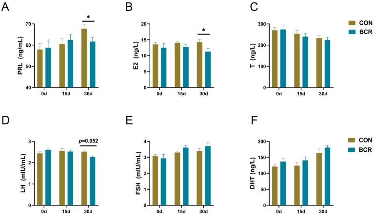
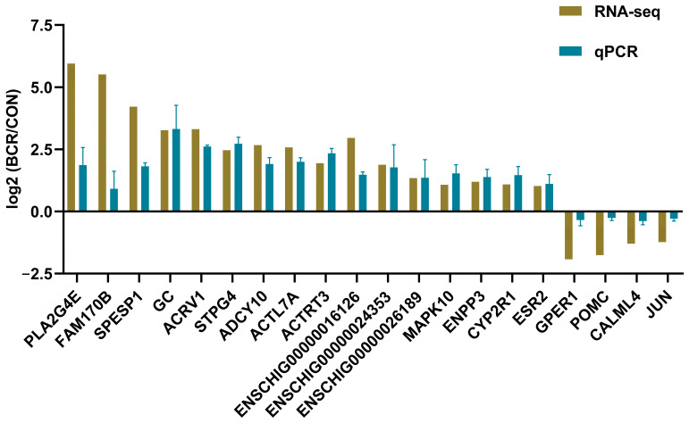
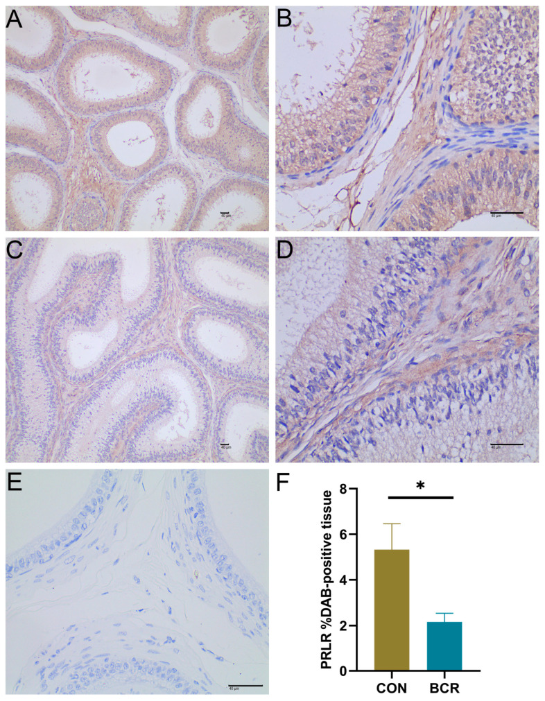
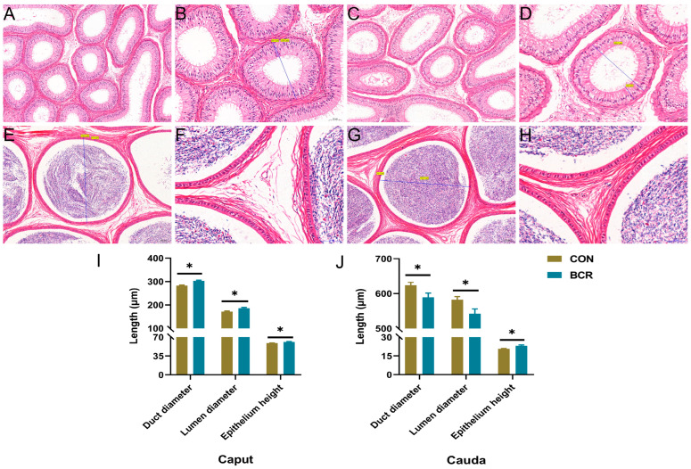
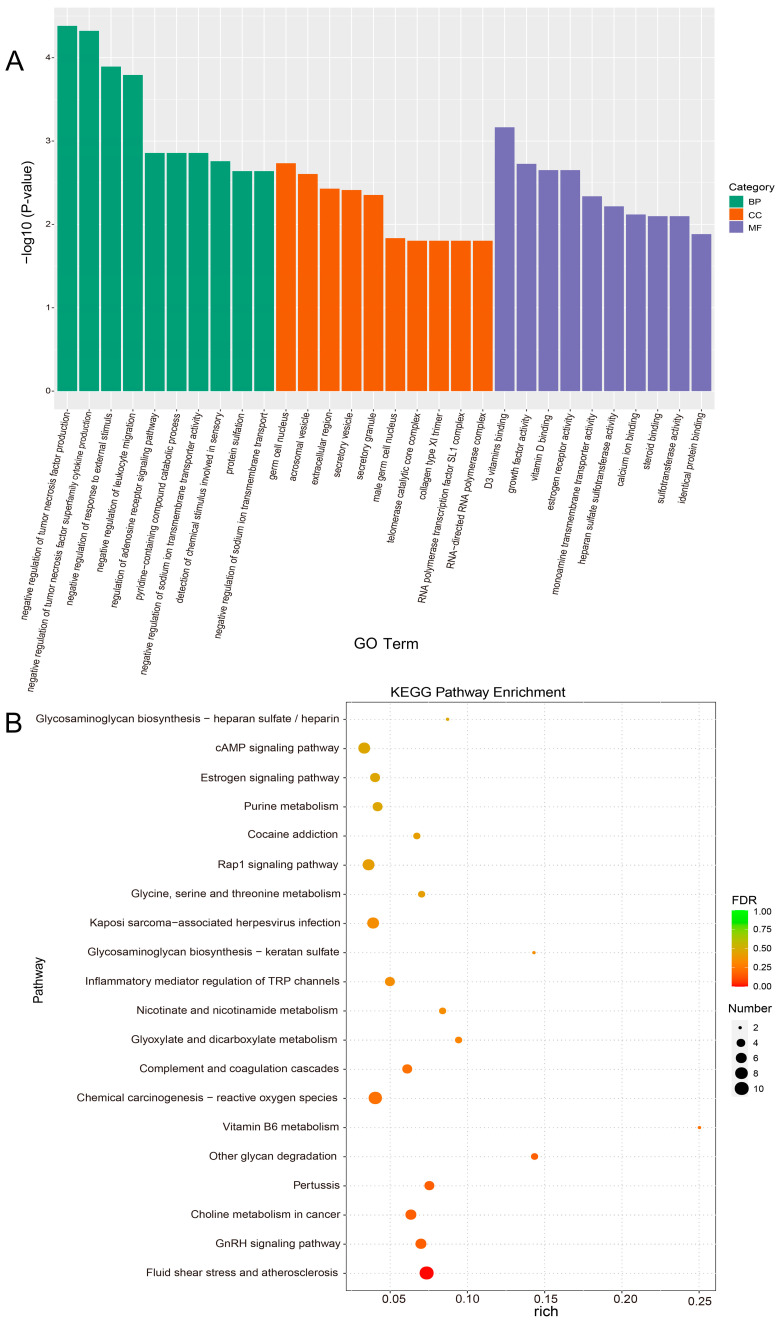
Yanshan Cashmere bucks are seasonal breeding animals and an important national genetic resource. This study aimed to investigate the involvement of prolactin (PRL) in the epididymal function of bucks. Twenty eleven-month-old Cashmere bucks were randomly divided into a control (CON) group and a bromocriptine (BCR, a prolactin inhibitor, 0.06 mg/kg body weight (BW)) treatment group. The experiment was conducted from September to October 2020 in Qinhuangdao City, China, and lasted for 30 days. Blood was collected on the last day before the BCR treatment (day 0) and on the 15th and 30th days after the BCR treatment (days 15 and 30). On the 30th day, all bucks were transported to the local slaughterhouse, where epididymal samples were collected immediately after slaughter. The left epididymis was preserved in 4% paraformaldehyde for histological observation, and the right epididymis was immediately preserved in liquid nitrogen for RNA sequencing (RNA-seq). The results show that the PRL inhibitor reduced the serum PRL and estradiol (E2) concentrations ( < 0.05) and tended to decrease luteinizing hormone (LH) concentrations ( = 0.052) by the 30th day, but no differences ( > 0.05) occurred by either day 0 or 15. There were no differences ( > 0.05) observed in the follicle-stimulating hormone (FSH), testosterone (T), and dihydrotestosterone (DHT) concentrations between the two groups. The PRL receptor (PRLR) protein was mainly located in the cytoplasm and intercellular substance of the epididymal epithelial cells. The PRL inhibitor decreased ( < 0.05) the expression of the PRLR protein in the epididymis. In the BCR group, the height of the epididymal epithelium in the caput and cauda increased, as did the diameter of the epididymal duct in the caput ( < 0.05). However, the diameter of the cauda epididymal duct decreased ( < 0.05). Thereafter, a total of 358 differentially expressed genes (DEGs) were identified in the epididymal tissues, among which 191 were upregulated and 167 were downregulated. Gene Ontology and Kyoto Encyclopedia of Genes and Genomes analyses revealed that , , , , and were mainly enriched in the estrogen signaling pathway, steroid binding, calcium ion binding, the GnRH signaling pathway, the cAMP signaling pathway, and the chemical carcinogenesis-reactive oxygen species pathway, which are related to epididymal function. In conclusion, the inhibition of PRL may affect the structure of the epididymis by reducing the expression of the PRLR protein and the secretion of E2. , , , , and could be the key genes of PRL in its regulation of epididymal reproductive function.
燕山绒山羊公羊是季节性繁殖动物,也是国家重要的遗传资源。本研究旨在探讨催乳素(PRL)在公羊附睾功能中的作用。将20只11月龄的绒山羊公羊随机分为对照组(CON)和溴隐亭(BCR,一种催乳素抑制剂,0.06 mg/kg体重(BW))处理组。实验于2020年9月至10月在中国秦皇岛市进行,为期30天。在BCR处理前的最后一天(第0天)以及BCR处理后的第15天和第30天(第15天和第30天)采集血液。在第30天,所有公羊被运至当地屠宰场,屠宰后立即采集附睾样本。左侧附睾保存在4%多聚甲醛中用于组织学观察,右侧附睾立即保存在液氮中用于RNA测序(RNA-seq)。结果表明,到第30天时,PRL抑制剂降低了血清PRL和雌二醇(E2)浓度(P<0.05),并使黄体生成素(LH)浓度有降低趋势(P = 0.052),但在第0天或第15天时无差异(P>0.05)。两组间促卵泡激素(FSH)、睾酮(T)和双氢睾酮(DHT)浓度无差异(P>0.05)。PRL受体(PRLR)蛋白主要位于附睾上皮细胞的细胞质和细胞间质中。PRL抑制剂降低了附睾中PRLR蛋白的表达(P<0.05)。在BCR组中,附睾头和附睾尾的上皮高度增加,附睾头的附睾管直径也增加(P<0.05)。然而,附睾尾的附睾管直径减小(P<0.05)。此后,在附睾组织中总共鉴定出358个差异表达基因(DEG),其中191个上调,167个下调。基因本体论和京都基因与基因组百科全书分析显示,……主要富集于雌激素信号通路、类固醇结合、钙离子结合、GnRH信号通路、cAMP信号通路和化学致癌-活性氧通路,这些通路与附睾功能相关。总之,抑制PRL可能通过降低PRLR蛋白表达和E2分泌来影响附睾结构。……可能是PRL调节附睾生殖功能的关键基因。