First Department of Comprehensive Internal Medicine of People's Hospital of Xinjiang Uygur Autonomous Region, No.91 Tianchi Road, Urumqi, 830001, Xinjiang, China.
Department of Urology, People's Hospital of Xinjiang Uygur Autonomous Region, No.91 Tianchi Road, Urumqi, 830001, Xinjiang, China.
Sci Rep. 2024 Jul 7;14(1):15635. doi: 10.1038/s41598-024-66433-y.
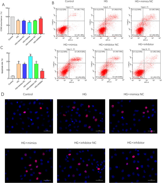
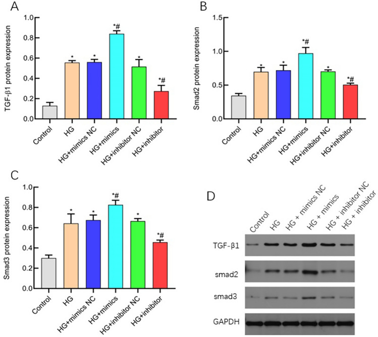
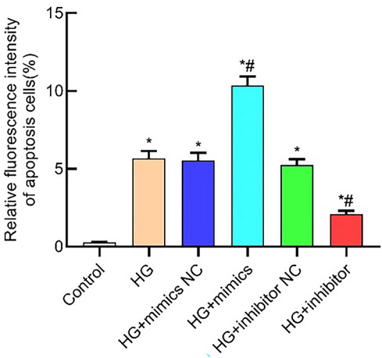
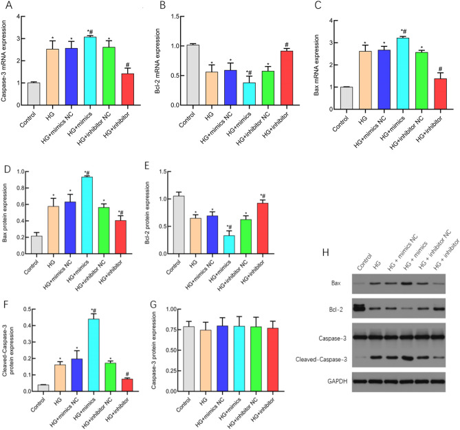
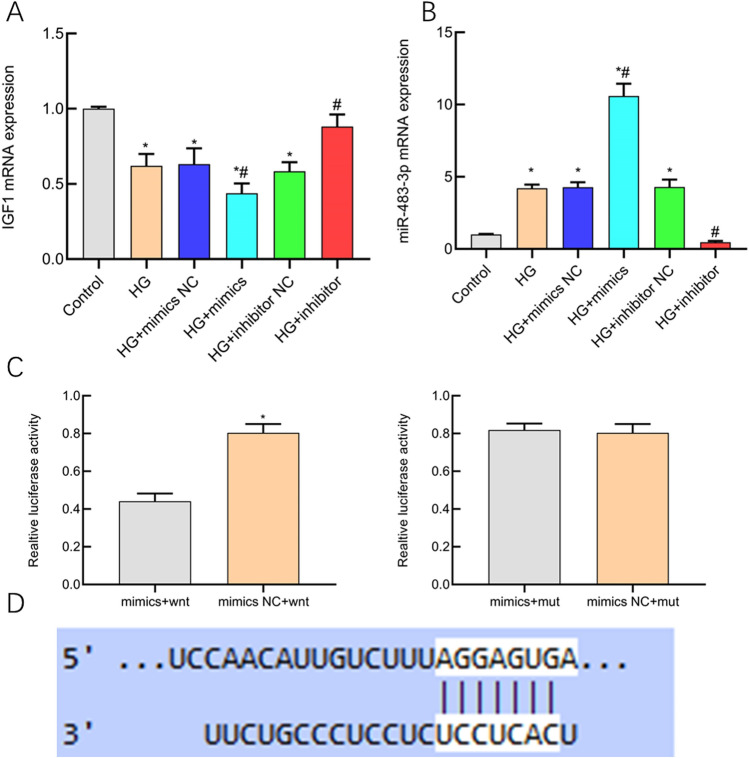
This study aimed to elucidate the influence of miR-483-3p on human renal tubular epithelial cells (HK-2) under high glucose conditions and to understand its mechanism. Human proximal tubular epithelial cells (HK-2) were exposed to 50 mmol/L glucose for 48 h to establish a renal tubular epithelial cell injury model, denoted as the high glucose group (HG group). Cells were also cultured for 48 h in a medium containing 5.5 mmol/L glucose, serving as the low glucose group. Transfection was performed in various groups: HK-2 + low glucose (control group), high glucose (50 mM) (HG group), high glucose + miR-483-3p mimics (HG + mimics group), high glucose +miR-483-3p inhibitor (HG + inhibitor group), and corresponding negative controls. Real-time quantitative polymerase chain reaction (qPCR) assessed the mRNA expression of miR-483-3p, bax, bcl-2, and caspase-3. Western blot determined the corresponding protein levels. Proliferation was assessed using the CCK-8 assay, and cell apoptosis was analyzed using the fluorescence TUNEL method. Western blot and Masson's staining were conducted to observe alterations in cell fibrosis post miR-483-3p transfection. Furthermore, a dual-luciferase assay investigated the targeting relationship between miR-483-3p and IGF-1. The CCK8 assay demonstrated that the HG + mimics group inhibited HK-2 cell proliferation, while the fluorescent TUNEL method revealed induced cell apoptosis in this group. Conversely, the HG + inhibitor group promoted cell proliferation and suppressed cell apoptosis. The HG + mimics group upregulated mRNA and protein expression of pro-apoptotic markers (bax and caspase-3), while downregulating anti-apoptotic marker (bcl-2) expression. In contrast, the HG + inhibitor group showed opposite effects. Collagen I and FN protein levels were significantly elevated in the HG + mimics group compared to controls (P < 0.05). Conversely, in the HG + inhibitor group, the protein expression of Collagen I and FN was notably reduced compared to the HG group (P < 0.05). The dual luciferase reporter assay confirmed that miR-483-3p could inhibit the luciferase activity of IGF-1's 3'-UTR region (P < 0.05). miR-483-3p exerts targeted regulation on IGF-1, promoting apoptosis and fibrosis in renal tubular epithelial cells induced by high glucose conditions.
本研究旨在阐明 miR-483-3p 在高糖环境下对人肾小管上皮细胞(HK-2)的影响,并探讨其机制。将人近端肾小管上皮细胞(HK-2)在 50mmol/L 葡萄糖中孵育 48h 建立肾小管上皮细胞损伤模型,记为高糖组(HG 组)。将细胞在含有 5.5mmol/L 葡萄糖的培养基中孵育 48h,作为低糖组。在以下各组中进行转染:HK-2+低糖(对照组)、高糖(50mM)(HG 组)、高糖+miR-483-3p 模拟物(HG+模拟物组)、高糖+miR-483-3p 抑制剂(HG+抑制剂组)和相应的阴性对照。实时定量聚合酶链反应(qPCR)检测 miR-483-3p、bax、bcl-2 和 caspase-3 的 mRNA 表达。Western blot 测定相应的蛋白水平。用 CCK-8 测定增殖,用荧光 TUNEL 法分析细胞凋亡。Western blot 和 Masson 染色观察 miR-483-3p 转染后细胞纤维化的变化。此外,双荧光素酶报告基因实验研究 miR-483-3p 与 IGF-1 之间的靶向关系。CCK8 检测结果表明,HG+模拟物组抑制 HK-2 细胞增殖,而荧光 TUNEL 法显示该组诱导细胞凋亡。相反,HG+抑制剂组促进细胞增殖并抑制细胞凋亡。HG+模拟物组上调促凋亡标志物(bax 和 caspase-3)的 mRNA 和蛋白表达,同时下调抗凋亡标志物(bcl-2)的表达。相反,HG+抑制剂组则表现出相反的效果。与对照组相比,HG+模拟物组的胶原 I 和 FN 蛋白水平显著升高(P<0.05)。相反,在 HG+抑制剂组中,与 HG 组相比,胶原 I 和 FN 的蛋白表达明显降低(P<0.05)。双荧光素酶报告基因实验证实,miR-483-3p 可以抑制 IGF-1 的 3'-UTR 区域的荧光素酶活性(P<0.05)。miR-483-3p 对 IGF-1 发挥靶向调节作用,促进高糖诱导的肾小管上皮细胞凋亡和纤维化。