Vue Zer, Murphy Alexandria, Le Han, Neikirk Kit, Garza-Lopez Edgar, Marshall Andrea G, Mungai Margaret, Jenkins Brenita, Vang Larry, Beasley Heather K, Ezedimma Mariaassumpta, Manus Sasha, Whiteside Aaron, Forni Maria Fernanda, Harris Chanel, Crabtree Amber, Albritton Claude F, Jamison Sydney, Demirci Mert, Prasad Praveena, Oliver Ashton, Actkins Ky'Era V, Shao Jianqiang, Zaganjor Elma, Scudese Estevão, Rodriguez Benjamin, Koh Alice, Rabago Izabella, Moore Johnathan E, Nguyen Desiree, Aftab Muhammad, Kirk Benjamin, Li Yahang, Wandira Nelson, Ahmad Taseer, Saleem Mohammad, Kadam Ashlesha, Katti Prasanna, Koh Ho-Jin, Evans Chantell, Koo Young Do, Wang Eric, Smith Quinton, Tomar Dhanendra, Williams Clintoria R, Sweetwyne Mariya T, Quintana Anita M, Phillips Mark A, Hubert David, Kirabo Annet, Dash Chandravanu, Jadiya Pooja, Kinder André, Ajijola Olujimi A, Miller-Fleming Tyne W, McReynolds Melanie R, Hinton Antentor
Department of Molecular Physiology and Biophysics, Vanderbilt University, Nashville, TN, 37232, USA.
Department of Biochemistry and Molecular Biology, The Huck Institute of the Life Sciences, Pennsylvania State University, State College, PA 16801.
bioRxiv. 2024 Jul 3:2024.06.20.599846. doi: 10.1101/2024.06.20.599846.
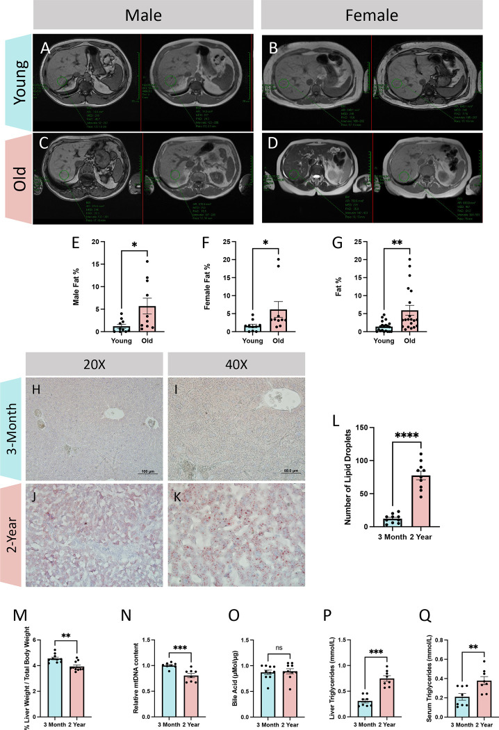
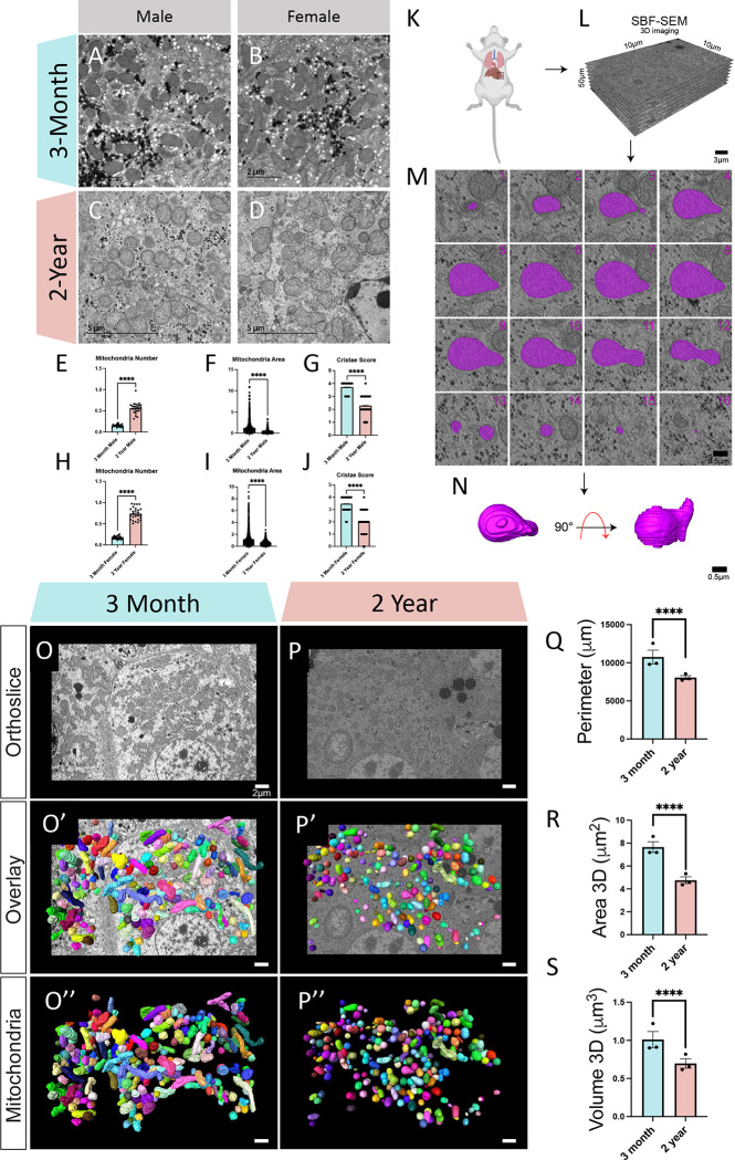
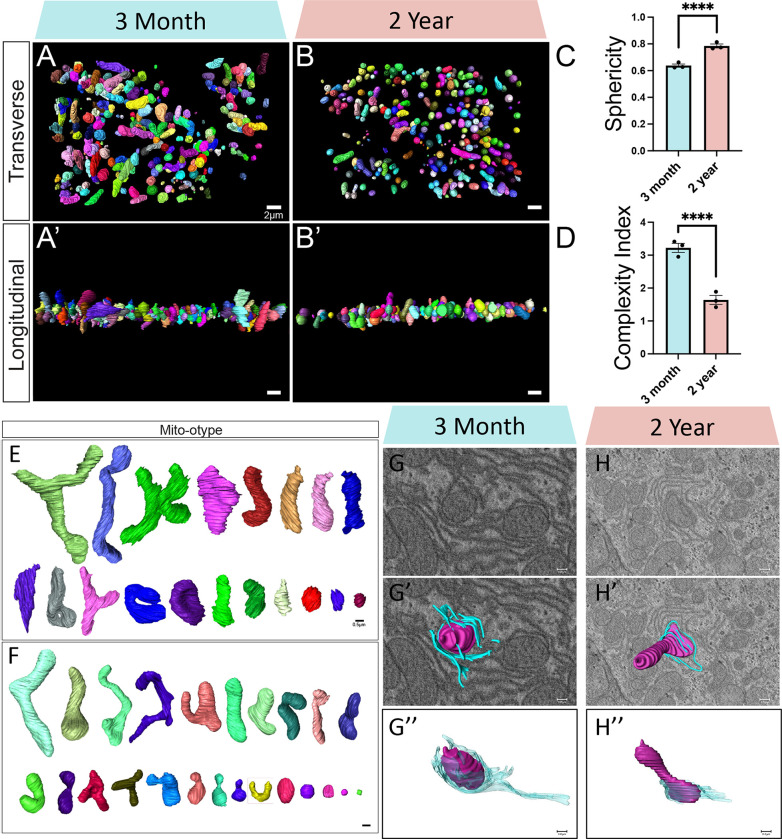
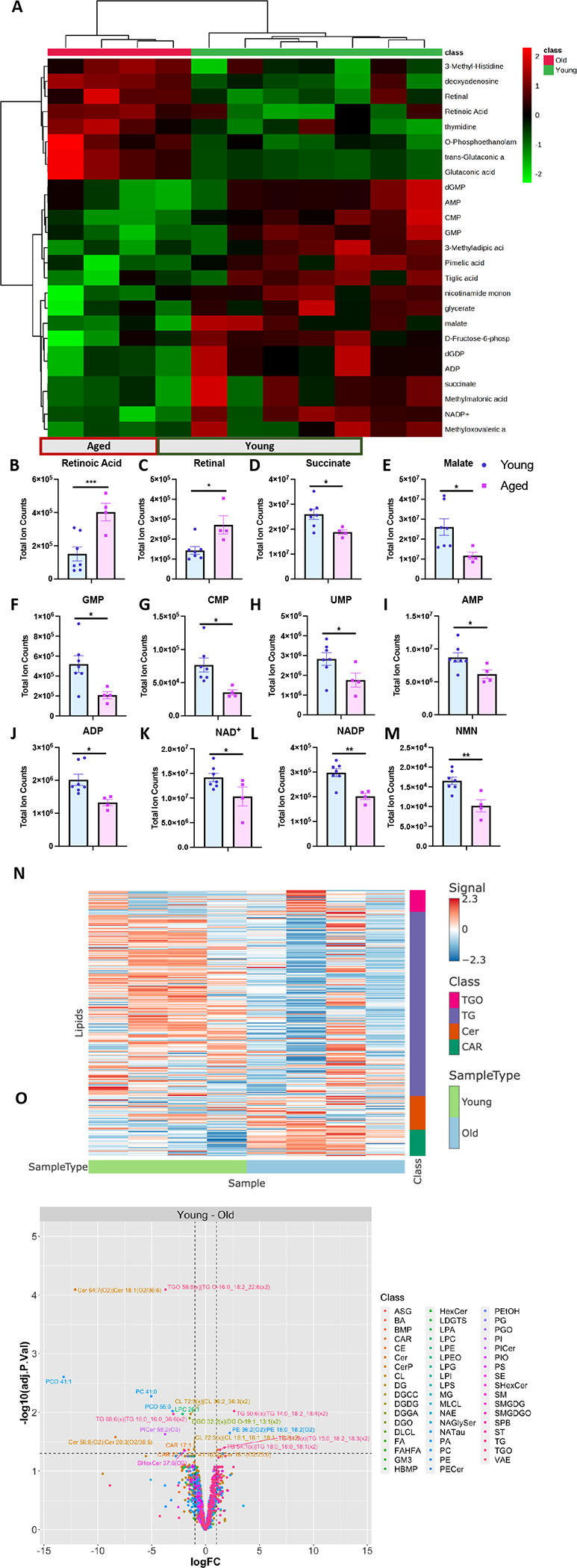
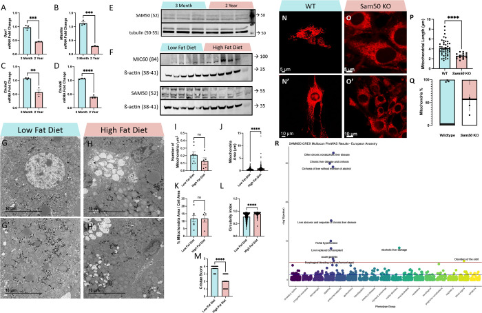
The liver, the largest internal organ and a metabolic hub, undergoes significant declines due to aging, affecting mitochondrial function and increasing the risk of systemic liver diseases. How the mitochondrial three-dimensional (3D) structure changes in the liver across aging, and the biological mechanisms regulating such changes confers remain unclear. In this study, we employed Serial Block Face-Scanning Electron Microscopy (SBF-SEM) to achieve high-resolution 3D reconstructions of murine liver mitochondria to observe diverse phenotypes and structural alterations that occur with age, marked by a reduction in size and complexity. We also show concomitant metabolomic and lipidomic changes in aged samples. Aged human samples reflected altered disease risk. To find potential regulators of this change, we examined the Mitochondrial Contact Site and Cristae Organizing System (MICOS) complex, which plays a crucial role in maintaining mitochondrial architecture. We observe that the MICOS complex is lost during aging, but not Sam50. Sam50 is a component of the sorting and assembly machinery (SAM) complex that acts in tandem with the MICOS complex to modulate cristae morphology. In murine models subjected to a high-fat diet, there is a marked depletion of the mitochondrial protein SAM50. This reduction in Sam50 expression may heighten the susceptibility to liver disease, as our human biobank studies corroborate that Sam50 plays a genetically regulated role in the predisposition to multiple liver diseases. We further show that changes in mitochondrial calcium dysregulation and oxidative stress accompany the disruption of the MICOS complex. Together, we establish that a decrease in mitochondrial complexity and dysregulated metabolism occur with murine liver aging. While these changes are partially be regulated by age-related loss of the MICOS complex, the confluence of a murine high-fat diet can also cause loss of Sam50, which contributes to liver diseases. In summary, our study reveals potential regulators that affect age-related changes in mitochondrial structure and metabolism, which can be targeted in future therapeutic techniques.
肝脏是最大的内部器官和代谢枢纽,由于衰老而出现显著衰退,影响线粒体功能并增加全身性肝脏疾病的风险。肝脏线粒体的三维(3D)结构在衰老过程中如何变化,以及调节这种变化的生物学机制仍不清楚。在本研究中,我们采用连续块面扫描电子显微镜(SBF-SEM)对小鼠肝脏线粒体进行高分辨率3D重建,以观察随年龄出现的各种表型和结构改变,其特征是大小和复杂性降低。我们还展示了老年样本中伴随的代谢组学和脂质组学变化。老年人类样本反映了疾病风险的改变。为了找到这种变化的潜在调节因子,我们研究了线粒体接触位点和嵴组织系统(MICOS)复合物,它在维持线粒体结构中起关键作用。我们观察到MICOS复合物在衰老过程中丢失,但Sam50没有。Sam50是分选和组装机器(SAM)复合物的一个组成部分,它与MICOS复合物协同作用以调节嵴的形态。在高脂饮食的小鼠模型中,线粒体蛋白SAM50明显减少。Sam50表达的这种降低可能会增加对肝脏疾病的易感性,因为我们的人类生物样本库研究证实Sam50在多种肝脏疾病的易感性中发挥基因调控作用。我们进一步表明,线粒体钙调节异常和氧化应激的变化伴随着MICOS复合物的破坏。总之,我们确定小鼠肝脏衰老会导致线粒体复杂性降低和代谢失调。虽然这些变化部分受与年龄相关的MICOS复合物缺失调节,但小鼠高脂饮食也会导致Sam50丢失,这会导致肝脏疾病。总之,我们的研究揭示了影响线粒体结构和代谢的年龄相关变化的潜在调节因子,这些因子可成为未来治疗技术的靶点。