• 文献检索
  • 文档翻译
  • 深度研究
  • 学术资讯
  • Suppr Zotero 插件Zotero 插件
  • 邀请有礼
  • 套餐&价格
  • 历史记录
应用&插件
Suppr Zotero 插件Zotero 插件浏览器插件Mac 客户端Windows 客户端微信小程序
定价
高级版会员购买积分包购买API积分包
服务
文献检索文档翻译深度研究API 文档MCP 服务
关于我们
关于 Suppr公司介绍联系我们用户协议隐私条款
关注我们

Suppr 超能文献

核心技术专利:CN118964589B侵权必究
粤ICP备2023148730 号-1Suppr @ 2026

文献检索

告别复杂PubMed语法,用中文像聊天一样搜索,搜遍4000万医学文献。AI智能推荐,让科研检索更轻松。

立即免费搜索

文件翻译

保留排版,准确专业,支持PDF/Word/PPT等文件格式,支持 12+语言互译。

免费翻译文档

深度研究

AI帮你快速写综述,25分钟生成高质量综述,智能提取关键信息,辅助科研写作。

立即免费体验

AGS蛋白中GoLoco基序的进化修饰有助于海胆胚胎中微细胞的形成。

The evolutionary modifications of a GoLoco motif in the AGS protein facilitate micromere formation in the sea urchin embryo.

作者信息

Emura Natsuko, Wavreil Florence D M, Fries Annaliese, Yajima Mamiko

机构信息

MCB Department, Brown University, 185 Meeting Street, BOX-GL277, Providence, RI 02912, USA.

出版信息

bioRxiv. 2024 Oct 15:2024.06.30.601440. doi: 10.1101/2024.06.30.601440.

DOI:10.1101/2024.06.30.601440
PMID:39005292
原文链接:https://pmc.ncbi.nlm.nih.gov/articles/PMC11244941/
Abstract

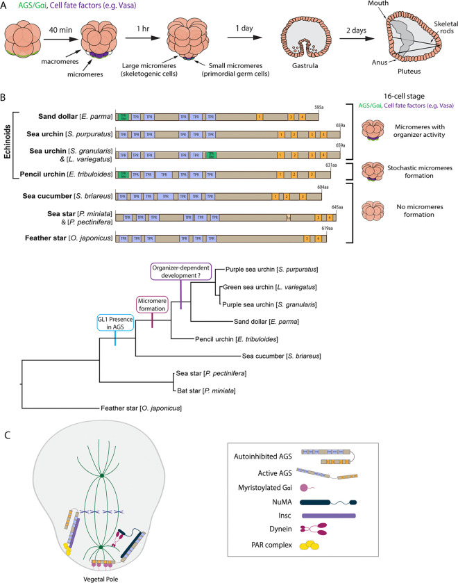
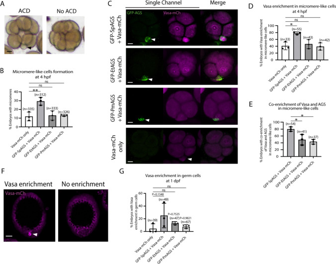
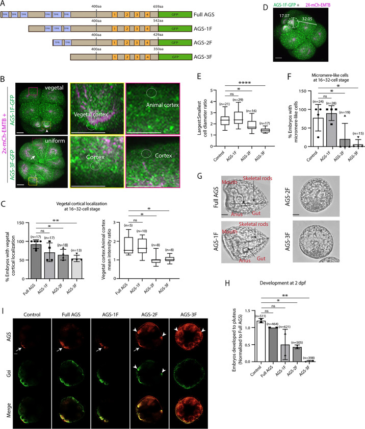
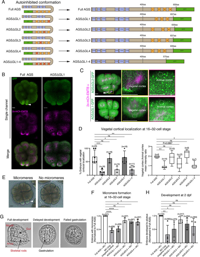
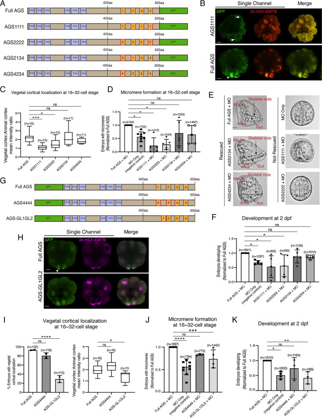
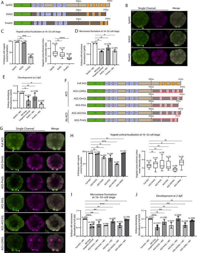
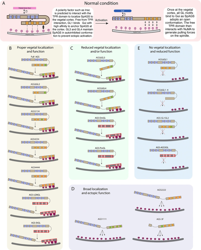
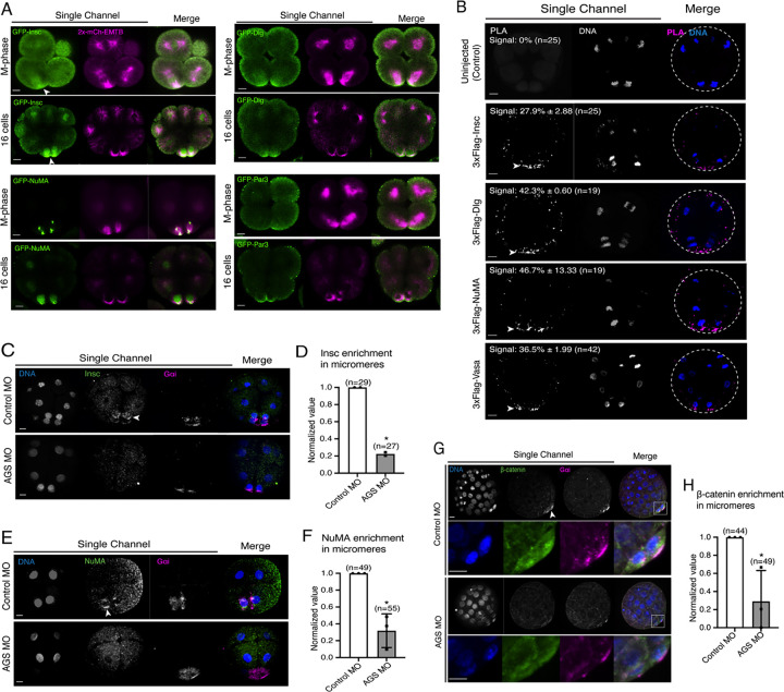
The evolutionary introduction of asymmetric cell division (ACD) into the developmental program facilitates the formation of a new cell type, contributing to developmental diversity and, eventually, to species diversification. The micromere of the sea urchin embryo may serve as one of those examples: An ACD at the 16-cell stage forms micromeres unique to echinoids among echinoderms. We previously reported that a polarity factor, Activator of G-protein Signaling (AGS), plays a crucial role in micromere formation. However, AGS and its associated ACD factors are present in all echinoderms and across most metazoans. This raises the question of what evolutionary modifications of AGS protein or its surrounding molecular environment contributed to the evolutionary acquisition of micromeres only in echinoids. In this study, we learned that the GoLoco motifs at the AGS C-terminus play critical roles in regulating micromere formation in sea urchin embryos. Further, other echinoderms' AGS or chimeric AGS that contain the C-terminus of AGS orthologs from various organisms showed varied localization and function in micromere formation. In contrast, the sea star or the pencil urchin orthologs of other ACD factors were consistently localized at the vegetal cortex in the sea urchin embryo, suggesting that AGS may be a unique variable factor that facilitates ACD diversity among echinoderms. Consistently, sea urchin AGS appears to facilitate micromere-like cell formation and accelerate the enrichment timing of the germline factor Vasa during early embryogenesis of the pencil urchin, an ancestral type of sea urchin. Based on these observations, we propose that the molecular evolution of a single polarity factor facilitates ACD diversity while preserving the core ACD machinery among echinoderms and beyond during evolution.

摘要

不对称细胞分裂(ACD)在发育程序中的进化引入促进了新细胞类型的形成,有助于发育多样性,并最终导致物种多样化。海胆胚胎的小分裂球可能就是其中一个例子:16细胞期的一次不对称细胞分裂形成了棘皮动物中独有的海胆类小分裂球。我们之前报道过,一种极性因子,G蛋白信号激活剂(AGS),在小分裂球形成中起关键作用。然而,AGS及其相关的不对称细胞分裂因子存在于所有棘皮动物以及大多数后生动物中。这就引出了一个问题,即AGS蛋白或其周围分子环境的哪些进化修饰导致了仅在海胆类中进化获得小分裂球。在这项研究中,我们发现AGS C末端的GoLoco基序在调节海胆胚胎小分裂球形成中起关键作用。此外,其他棘皮动物的AGS或包含来自各种生物的AGS直系同源物C末端的嵌合AGS在小分裂球形成中表现出不同的定位和功能。相比之下,其他不对称细胞分裂因子的海星或铅笔海胆直系同源物在海胆胚胎中始终定位于植物皮层,这表明AGS可能是促进棘皮动物间不对称细胞分裂多样性的独特可变因素。一致地,海胆AGS似乎促进了类小分裂球细胞的形成,并加速了铅笔海胆(海胆的一种原始类型)早期胚胎发育过程中生殖系因子Vasa的富集时间。基于这些观察结果,我们提出单个极性因子的分子进化促进了不对称细胞分裂多样性,同时在进化过程中保留了棘皮动物及其他生物中的核心不对称细胞分裂机制。

https://cdn.ncbi.nlm.nih.gov/pmc/blobs/e7f6/11488036/3a6c66375c92/nihpp-2024.06.30.601440v2-f0010.jpg
https://cdn.ncbi.nlm.nih.gov/pmc/blobs/e7f6/11488036/61b3150c0901/nihpp-2024.06.30.601440v2-f0001.jpg
https://cdn.ncbi.nlm.nih.gov/pmc/blobs/e7f6/11488036/45b2aef96e85/nihpp-2024.06.30.601440v2-f0002.jpg
https://cdn.ncbi.nlm.nih.gov/pmc/blobs/e7f6/11488036/c8e3b662bfac/nihpp-2024.06.30.601440v2-f0003.jpg
https://cdn.ncbi.nlm.nih.gov/pmc/blobs/e7f6/11488036/328efef0235f/nihpp-2024.06.30.601440v2-f0004.jpg
https://cdn.ncbi.nlm.nih.gov/pmc/blobs/e7f6/11488036/ccf82b3e3d24/nihpp-2024.06.30.601440v2-f0005.jpg
https://cdn.ncbi.nlm.nih.gov/pmc/blobs/e7f6/11488036/4c714b716f81/nihpp-2024.06.30.601440v2-f0006.jpg
https://cdn.ncbi.nlm.nih.gov/pmc/blobs/e7f6/11488036/3476d40dad89/nihpp-2024.06.30.601440v2-f0007.jpg
https://cdn.ncbi.nlm.nih.gov/pmc/blobs/e7f6/11488036/11aeda72b1b9/nihpp-2024.06.30.601440v2-f0008.jpg
https://cdn.ncbi.nlm.nih.gov/pmc/blobs/e7f6/11488036/b79aabeb29c7/nihpp-2024.06.30.601440v2-f0009.jpg
https://cdn.ncbi.nlm.nih.gov/pmc/blobs/e7f6/11488036/3a6c66375c92/nihpp-2024.06.30.601440v2-f0010.jpg

相似文献

1
The evolutionary modifications of a GoLoco motif in the AGS protein facilitate micromere formation in the sea urchin embryo.AGS蛋白中GoLoco基序的进化修饰有助于海胆胚胎中微细胞的形成。
bioRxiv. 2024 Oct 15:2024.06.30.601440. doi: 10.1101/2024.06.30.601440.
2
The evolutionary modifications of a GoLoco motif in the AGS protein facilitate micromere formation in the sea urchin embryo.AGS蛋白中GoLoco基序的进化修饰促进了海胆胚胎中微小分裂球的形成。
Elife. 2024 Dec 23;13:RP100086. doi: 10.7554/eLife.100086.
3
Evolutionary modification of AGS protein contributes to formation of micromeres in sea urchins.AGS 蛋白的进化修饰有助于海胆形成小分裂球。
Nat Commun. 2019 Aug 22;10(1):3779. doi: 10.1038/s41467-019-11560-8.
4
Micromere formation and its evolutionary implications in the sea urchin.微小分裂的形成及其在海胆中的进化意义。
Curr Top Dev Biol. 2022;146:211-238. doi: 10.1016/bs.ctdb.2021.10.008. Epub 2021 Dec 3.
5
An evolutionary transition of Vasa regulation in echinoderms.棘皮动物中瓦萨基因调控的进化转变。
Evol Dev. 2009 Sep-Oct;11(5):560-73. doi: 10.1111/j.1525-142X.2009.00362.x.
6
Activator of G-protein signaling in asymmetric cell divisions of the sea urchin embryo.海胆胚胎不对称细胞分裂中G蛋白信号通路激活剂。
Dev Growth Differ. 2006 Dec;48(9):549-57. doi: 10.1111/j.1440-169X.2006.00895.x.
7
Nuclear beta-catenin is required to specify vegetal cell fates in the sea urchin embryo.核β-连环蛋白是海胆胚胎中确定植物细胞命运所必需的。
Development. 1999 Jan;126(2):345-57. doi: 10.1242/dev.126.2.345.
8
Micromere descendants at the blastula stage are involved in normal archenteron formation in sea urchin embryos.囊胚期的小分裂球后代参与海胆胚胎正常原肠的形成。
Dev Genes Evol. 2001 Feb;211(2):83-8. doi: 10.1007/s004270000120.
9
Effect on Inhibition of Micromere Segregation on the Mitotic Pattern in the Sea Urchin Embryo: (micromeres/mitotic pattern/sea urchin).对海胆胚胎有丝分裂模式中微裂球分离抑制的影响:(微裂球/有丝分裂模式/海胆)
Dev Growth Differ. 1985;27(1):29-34. doi: 10.1111/j.1440-169X.1985.00029.x.
10
The micro1 gene is necessary and sufficient for micromere differentiation and mid/hindgut-inducing activity in the sea urchin embryo.微1基因对于海胆胚胎中的小分裂球分化和中/后肠诱导活性而言是必需且充分的。
Dev Genes Evol. 2005 Sep;215(9):450-59. doi: 10.1007/s00427-005-0006-y. Epub 2005 Aug 3.