Daga Kanupriya R, Larey Andrew M, Morfin Maria G, Chen Kailin, Bitarafan Sara, Carpenter Jana M, Hynds Hannah M, Hines Kelly M, Wood Levi B, Marklein Ross A
School of Chemical, Materials, and Biomedical Engineering, University of Georgia, Athens, GA, USA.
Regenerative Bioscience Center, University of Georgia, Athens, GA, USA.
bioRxiv. 2024 Jul 3:2024.07.01.601612. doi: 10.1101/2024.07.01.601612.
Mesenchymal stromal cell derived extracellular vesicles (MSC-EVs) are a promising therapeutic for neuroinflammation. MSC-EVs can interact with microglia, the resident immune cells of the brain, to exert their immunomodulatory effects. In response to inflammatory cues, such as cytokines, microglia undergo phenotypic changes indicative of their function e.g. morphology and secretion. However, these changes in response to MSC-EVs are not well understood. Additionally, no disease-relevant screening tools to assess MSC-EV bioactivity exist, which has further impeded clinical translation. Here, we developed a quantitative, high throughput morphological profiling approach to assess the response of microglia to neuroinflammation-relevant signals and whether this morphological response can be used to indicate the bioactivity of MSC-EVs.
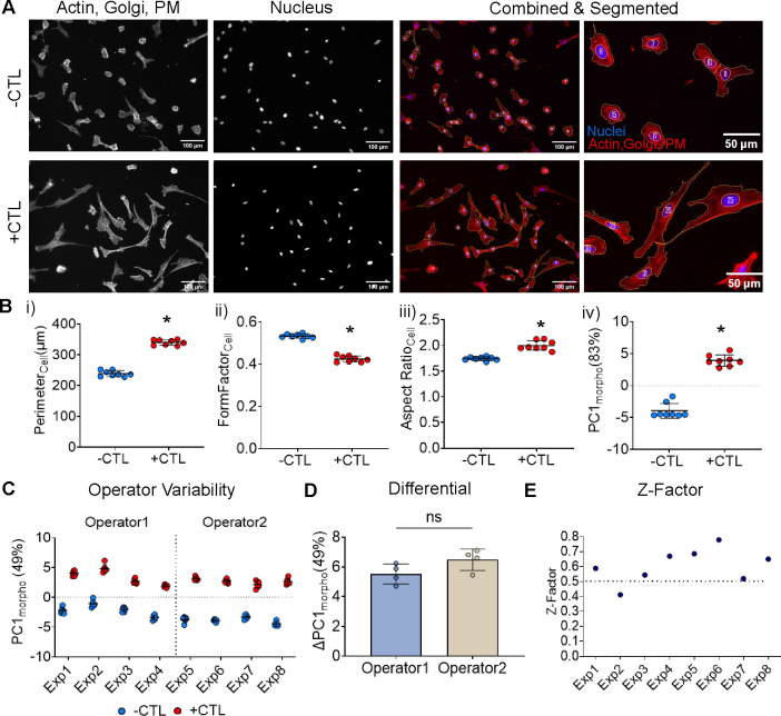
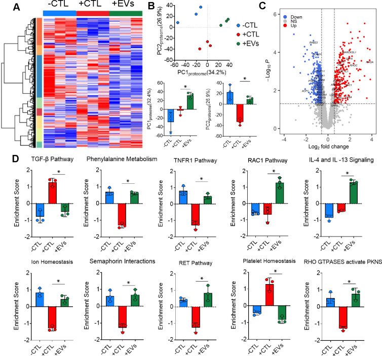
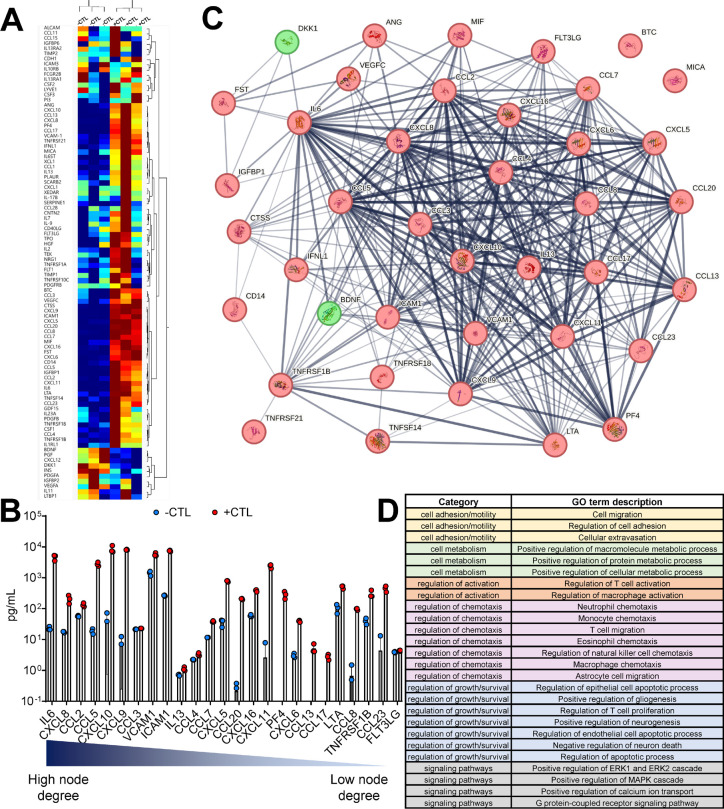
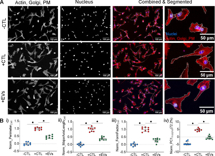
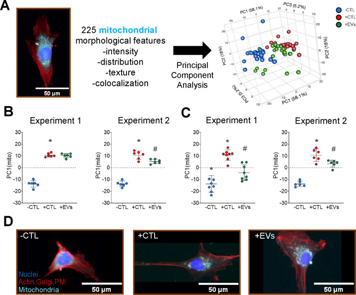
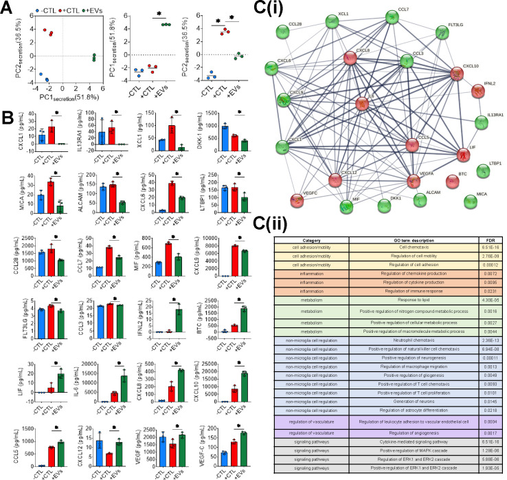
Using an immortalized human microglia cell-line, we observed increased size (perimeter, major axis length) and complexity (form factor) upon stimulation with interferon-gamma (IFN-γ) and tumor necrosis factor-alpha (TNF-α). Upon treatment with MSC-EVs, the overall morphological score (determined using principal component analysis) shifted towards the unstimulated morphology, indicating that MSC-EVs are bioactive and modulate microglia. The morphological effects of MSC-EVs in TNF-γ/IFN-α stimulated cells were concomitant with reduced secretion of 14 chemokines/cytokines (e.g. CXCL6, CXCL9) and increased secretion of 12 chemokines/cytokines (e.g. CXCL8, CXCL10). Proteomic analysis of cell lysates revealed significant increases in 192 proteins (e.g. HIBADH, MEAK7, LAMC1) and decreases in 257 proteins (e.g. PTEN, TOM1, MFF) with MSC-EV treatment. Of note, many of these proteins are involved in regulation of cell morphology and migration. Gene Set Variation Analysis revealed upregulation of pathways associated with immune response, such as regulation of cytokine production, immune cell infiltration (e.g. T cells, NK cells) and morphological changes (e.g. Semaphorin, RHO/Rac signaling). Additionally, changes in microglia mitochondrial morphology were measured suggesting that MSC-EV modulate mitochondrial metabolism.
This study comprehensively demonstrates the effects of MSC-EVs on human microglial morphology, cytokine secretion, cellular proteome, and mitochondrial content. Our high-throughput, rapid, low-cost morphological approach enables screening of MSC-EV batches and manufacturing conditions to enhance EV function and mitigate EV functional heterogeneity in a disease relevant manner. This approach is highly generalizable and can be further adapted and refined based on selection of the disease-relevant signal, target cell, and therapeutic product.
间充质基质细胞衍生的细胞外囊泡(MSC-EVs)是一种很有前景的神经炎症治疗方法。MSC-EVs可与脑内常驻免疫细胞小胶质细胞相互作用,发挥其免疫调节作用。响应细胞因子等炎症信号时,小胶质细胞会发生表型变化,这些变化反映了它们的功能,如形态和分泌情况。然而,对MSC-EVs引起的这些变化了解并不充分。此外,目前尚无评估MSC-EV生物活性的疾病相关筛选工具,这进一步阻碍了其临床转化。在此,我们开发了一种定量、高通量的形态学分析方法,以评估小胶质细胞对神经炎症相关信号的反应,以及这种形态学反应是否可用于指示MSC-EVs的生物活性。
使用永生化的人小胶质细胞系,我们观察到在用干扰素-γ(IFN-γ)和肿瘤坏死因子-α(TNF-α)刺激后,细胞大小(周长、长轴长度)和复杂性(形状因子)增加。在用MSC-EVs处理后,总体形态学评分(使用主成分分析确定)向未刺激的形态转变,表明MSC-EVs具有生物活性并可调节小胶质细胞。MSC-EVs对TNF-γ/IFN-α刺激细胞的形态学影响伴随着14种趋化因子/细胞因子(如CXCL6、CXCL9)分泌减少和12种趋化因子/细胞因子(如CXCL8、CXCL10)分泌增加。对细胞裂解物的蛋白质组学分析显示,经MSC-EV处理后,192种蛋白质(如HIBADH、MEAK7、LAMC1)显著增加,257种蛋白质(如PTEN、TOM1、MFF)减少。值得注意的是,其中许多蛋白质参与细胞形态和迁移的调节。基因集变异分析显示与免疫反应相关的通路上调,如细胞因子产生的调节、免疫细胞浸润(如T细胞、NK细胞)和形态变化(如信号素、RHO/Rac信号)。此外,还测量了小胶质细胞线粒体形态的变化,表明MSC-EV可调节线粒体代谢。
本研究全面证明了MSC-EVs对人小胶质细胞形态、细胞因子分泌、细胞蛋白质组和线粒体含量的影响。我们的高通量、快速、低成本的形态学方法能够筛选MSC-EV批次和制造条件,以以与疾病相关的方式增强EV功能并减轻EV功能异质性。这种方法具有高度的通用性,可根据疾病相关信号、靶细胞和治疗产品的选择进一步调整和完善。