Department of Orbit, Oculoplasty, Aesthetic and Reconstructive Services, Sankara Nethralaya, Medical Research Foundation, Chennai, Tamil Nadu, India.
Department of Radiology, Sankara Nethralaya, Medical Research Foundation, Chennai, Tamil Nadu, India.
Middle East Afr J Ophthalmol. 2024 Jun 14;30(2):98-102. doi: 10.4103/meajo.meajo_7_23. eCollection 2023 Apr-Jun.
The objective is to analyze the radiological diagnosis of orbital lesions and their correlation with the final histopathological findings. We compared the initial reports by extramural radiologists and an in-house radiologist specialized in orbital imaging to evaluate the diagnostic accuracy in the interpretation of orbital imaging.
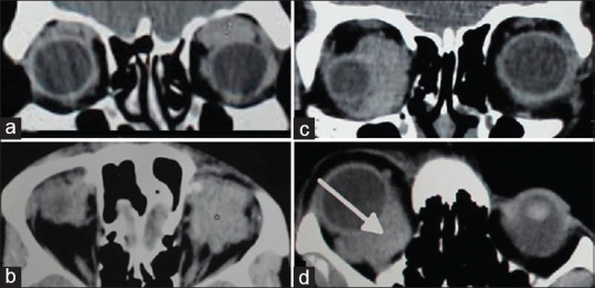
This was a retrospective chart review of forty patients referred to a Tertiary Eye Care Center in South India over a period of 7 years. These patients already had their imaging done elsewhere. The imaging was re-evaluated by an in-house radiologist. The radiological findings were correlated with the histopathological diagnosis. The diagnostic accuracy between the two radiologists was compared. The sensitivity, specificity, positive predictive value (PPV), and negative predictive value in differentiating malignant from benign lesions were calculated in both groups. The sensitivity and PPV of the radiological diagnosis for neoplastic and inflammatory lesions in both groups were analyzed.
The accuracy in differentiating malignant from benign and inflammatory lesions by our in-house radiologist and extramural radiologists was 95% (κ = 0.9 [0.764, 0.997]) and 50% (κ = 0.036 [-0.160, 0.232]), respectively. The sensitivity and PPV of the radiological diagnosis by our in-house radiologist were 93.31% and 100% for benign lesions and 95.24% and 95.24% for malignant lesions. On the contrary, reports from the extramural radiologists showed a sensitivity and PPV of 76.92% and 66.67% for benign lesions and 14.28% and 60% for malignant lesions.
A high radiological diagnostic accuracy is possible when analyzed by radiologists experienced in orbital imaging.
分析眼眶病变的放射诊断及其与最终组织病理学发现的相关性。我们比较了外部放射科医生和专门从事眼眶成像的内部放射科医生的初始报告,以评估在解释眼眶成像方面的诊断准确性。
这是一项对印度南部一家三级眼科护理中心的 40 名患者进行的回顾性图表审查,这些患者已经在其他地方进行了影像学检查。对内部放射科医生重新评估了这些图像。将放射学发现与组织病理学诊断进行了比较。比较了两位放射科医生的诊断准确性。计算了两组中恶性与良性病变、炎症病变的敏感性、特异性、阳性预测值(PPV)和阴性预测值。分析了两组中放射诊断对肿瘤和炎症病变的敏感性和 PPV。
我们的内部放射科医生和外部放射科医生区分恶性与良性和炎症病变的准确性分别为 95%(κ=0.9[0.764,0.997])和 50%(κ=0.036[-0.160,0.232])。内部放射科医生的放射诊断的敏感性和 PPV 分别为 93.31%和 100%用于良性病变,95.24%和 95.24%用于恶性病变。相比之下,外部放射科医生的报告显示,良性病变的敏感性和 PPV 分别为 76.92%和 66.67%,恶性病变的敏感性和 PPV 分别为 14.28%和 60%。
当由具有丰富眼眶成像经验的放射科医生进行分析时,放射学诊断的准确性较高。