Amland Rachel, Selbæk Geir, Brækhus Anne, Edwin Trine H, Engedal Knut, Knapskog Anne-Brita, Olsrud Ellen Regine, Persson Karin
The Norwegian National Centre for Ageing and Health, Vestfold Hospital Trust, Tønsberg, Norway.
Department of Geriatric Medicine, Oslo University Hospital, Oslo, Norway.
Front Neurol. 2024 Jul 1;15:1425502. doi: 10.3389/fneur.2024.1425502. eCollection 2024.
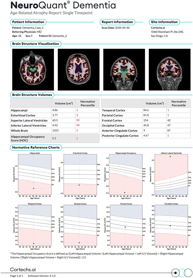
BACKGROUND/AIMS: The number of patients suffering from cognitive decline and dementia increases, and new possible treatments are being developed. Thus, the need for time efficient and cost-effective methods to facilitate an early diagnosis and prediction of future cognitive decline in patients with early cognitive symptoms is becoming increasingly important. The aim of this study was to evaluate whether an MRI based software, NeuroQuant® (NQ), producing volumetry of the hippocampus and whole brain volume (WBV) could predict: (1) conversion from subjective cognitive decline (SCD) at baseline to mild cognitive impairment (MCI) or dementia at follow-up, and from MCI at baseline to dementia at follow-up and (2) progression of cognitive and functional decline defined as an annual increase in the Clinical Dementia Rating Scale Sum of Boxes (CDR-SB) score.
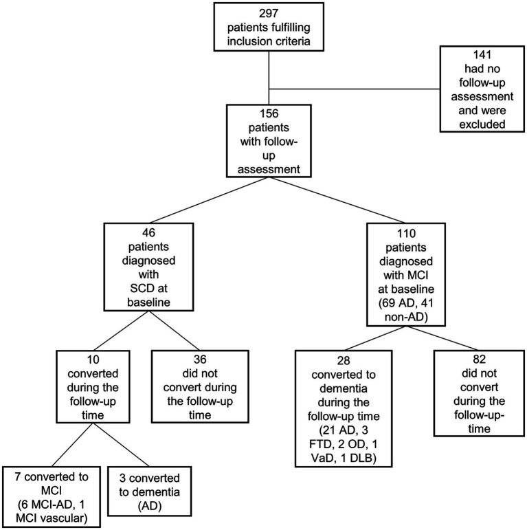
MRI was performed in 156 patients with SCD or MCI from the memory clinic at Oslo University Hospital (OUH) that had been assessed with NQ and had a clinical follow-up examination. Logistic and linear regression analyses were performed with hippocampus volume and WBV as independent variables, and conversion or progression as dependent variables, adjusting for demographic and other relevant covariates including Mini-Mental State Examination-Norwegian Revised Version score (MMSE-NR) and Apolipoprotein E ɛ4 ( ɛ4) carrier status.
Hippocampus volume, but not WBV, was associated with conversion to MCI or dementia, but neither were associated with conversion when adjusting for MMSE-NR. Both hippocampus volume and WBV were associated with progression as measured by the annual change in CDR-SB score in both unadjusted and adjusted analyses.
The results indicate that automated regional MRI volumetry of the hippocampus and WBV can be useful in predicting further cognitive decline in patients with early cognitive symptoms.
背景/目的:认知功能减退和痴呆患者的数量不断增加,新的潜在治疗方法也在不断研发。因此,对于采用高效且经济的方法来促进对有早期认知症状患者的未来认知功能减退进行早期诊断和预测的需求日益重要。本研究的目的是评估一款基于磁共振成像(MRI)的软件NeuroQuant®(NQ),其可生成海马体积和全脑体积(WBV),能否预测:(1)从基线时的主观认知功能减退(SCD)转变为随访时的轻度认知障碍(MCI)或痴呆,以及从基线时的MCI转变为随访时的痴呆;(2)认知和功能减退的进展,定义为临床痴呆评定量表总盒数(CDR-SB)得分的年度增加。
对奥斯陆大学医院(OUH)记忆门诊的156例SCD或MCI患者进行了MRI检查,这些患者已接受NQ评估并进行了临床随访检查。以海马体积和WBV作为自变量,以转变或进展作为因变量进行逻辑回归和线性回归分析,并对人口统计学和其他相关协变量进行校正,包括简易精神状态检查表挪威修订版得分(MMSE-NR)和载脂蛋白E ε4(ε4)携带状态。
海马体积而非WBV与转变为MCI或痴呆相关,但在校正MMSE-NR后两者均与转变无关。在未校正和校正分析中,海马体积和WBV均与以CDR-SB得分年度变化衡量的进展相关。
结果表明,海马和WBV的自动化MRI区域体积测量可用于预测有早期认知症状患者的进一步认知功能减退。