Tsui Jonathan C, Aleman Tomas S, Tapino Paul J, Kim Benjamin J
Scheie Eye Institute, Perelman School of Medicine, University of Pennsylvania, Philadelphia, PA, USA.
Department of Ophthalmology, Veterans Affairs New Jersey Healthcare System, East Orange, NJ, USA.
Case Rep Ophthalmol. 2024 Jun 12;15(1):497-506. doi: 10.1159/000538045. eCollection 2024 Jan-Dec.
We report a case of pseudoxanthoma elasticum (PXE) with an atypical phenotype likely related to a hypomorphic variant in .
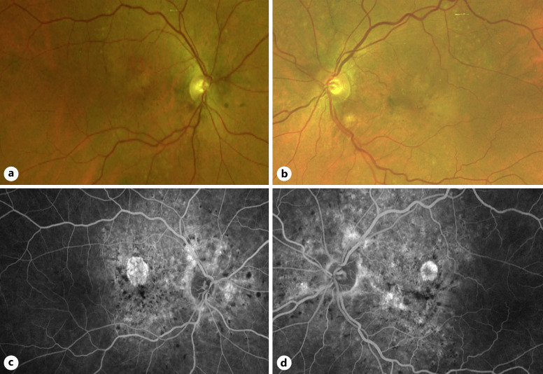
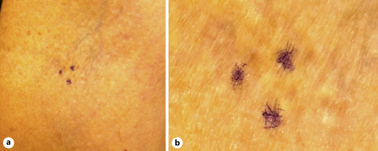
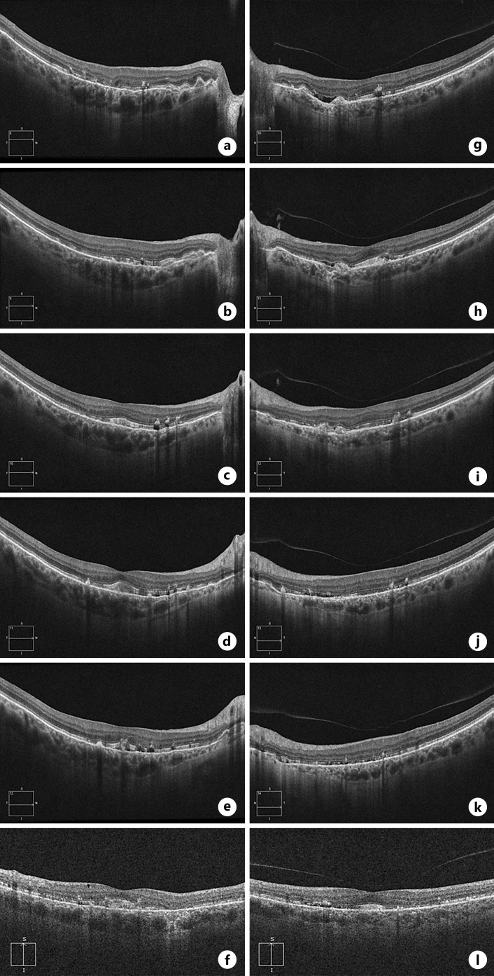
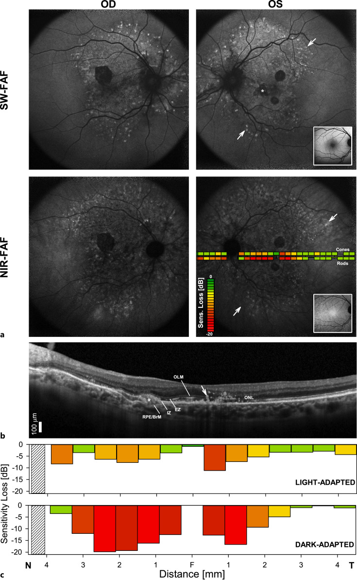
A 66-year-old Caucasian female with a history of a maculopathy interpreted as either age-related macular degeneration or a pattern dystrophy underwent a detailed ophthalmic evaluation. Visual acuities were 20/25, OD, and 20/20, OS. Spectral domain optical coherence and fluorescein angiography demonstrated outer retinal disruptions and breaks in retinal pigment epithelium (RPE)/Bruch's membrane bilaterally, consistent with angioid streaks. A large area of hypo- and hyperautofluorescence extending from the central retina into the peripapillary retina was documented with short-wavelength excitation autofluorescence. The area of hypoautofluorescence, which was much larger on near-infrared excitation, spared the temporal retina. Two-color dark-adapted perimetries documented severe rod sensitivity losses and less severe cone sensitivity abnormalities co-localizing with the RPE abnormalities. No obvious skin findings were observed, and initial dermatologic biopsy was negative. Gene screening identified a pathogenic gene variant c.1552C>T and a previously reported variant of uncertain significance c.1171A>G. A second dermatologic biopsy demonstrated positive findings consistent with PXE.
Although this patient had minimal skin findings, this patient had characteristic structural and functional abnormalities of a pattern dystrophy with angioid streaks and histologic evidence of PXE, suggesting compound heterozygous variants involving the hypomorphic ABCC6 c.1171A>G variant. These findings support the pathogenic role of both variants.
我们报告一例弹性假黄瘤(PXE),其非典型表型可能与某个基因的低表达变异有关。
一名66岁的白种女性,有黄斑病变史,曾被诊断为年龄相关性黄斑变性或图案状营养不良,接受了详细的眼科评估。右眼视力为20/25,左眼视力为20/20。频域光学相干断层扫描和荧光素血管造影显示双侧视网膜外层破坏以及视网膜色素上皮(RPE)/布鲁赫膜破裂,符合血管样条纹表现。短波长激发自发荧光记录到从中央视网膜延伸至视乳头周围视网膜的大片低自发荧光和高自发荧光区域。近红外激发下低自发荧光区域更大,颞侧视网膜未受累。双色暗适应视野检查记录到严重的视杆细胞敏感性丧失以及与RPE异常共定位的较轻的视锥细胞敏感性异常。未观察到明显的皮肤表现,初次皮肤活检结果为阴性。基因筛查发现一个致病基因变异c.1552C>T和一个先前报道的意义不确定的变异c.1171A>G。第二次皮肤活检显示与PXE一致的阳性结果。
尽管该患者皮肤表现轻微,但具有图案状营养不良伴血管样条纹的特征性结构和功能异常以及PXE的组织学证据,提示涉及低表达ABCC6 c.1171A>G变异的复合杂合变异。这些发现支持这两种变异的致病作用。