Department of Biophysics and Radiation Biology, Semmelweis University, Tűzoltó utca 37-47, 1094, Budapest, Hungary.
In Vivo Imaging Advanced Core Facility, Hungarian Centre of Excellence for Molecular Medicine, Tűzoltó utca 37-47, 1094, Budapest, Hungary.
Cell Mol Biol Lett. 2024 Jul 19;29(1):105. doi: 10.1186/s11658-024-00619-0.
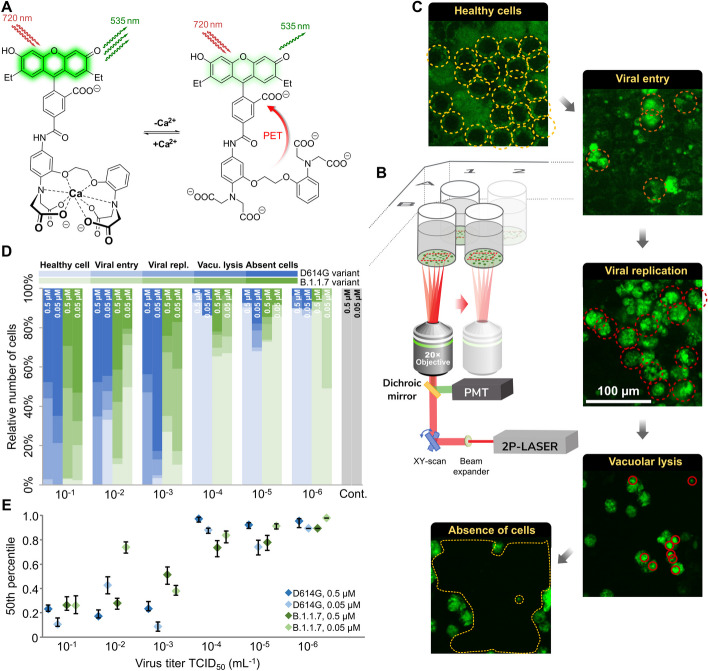
The organism-wide effects of severe acute respiratory syndrome coronavirus 2 (SARS-CoV-2) viral infection are well studied, but little is known about the dynamics of how the infection spreads in time among or within cells due to the scarcity of suitable high-resolution experimental systems. It has been reported that SARS-CoV-2 infection pathways converge at calcium influx and subcellular calcium distribution changes. Imaging combined with a proper staining technique is an effective tool for studying subcellular calcium-related infection and replication mechanisms at such resolutions.
Using two-photon (2P) fluorescence imaging with our novel Ca-selective dye, automated image analysis and clustering analysis were applied to reveal titer and variant effects on SARS-CoV-2-infected Vero E6 cells.
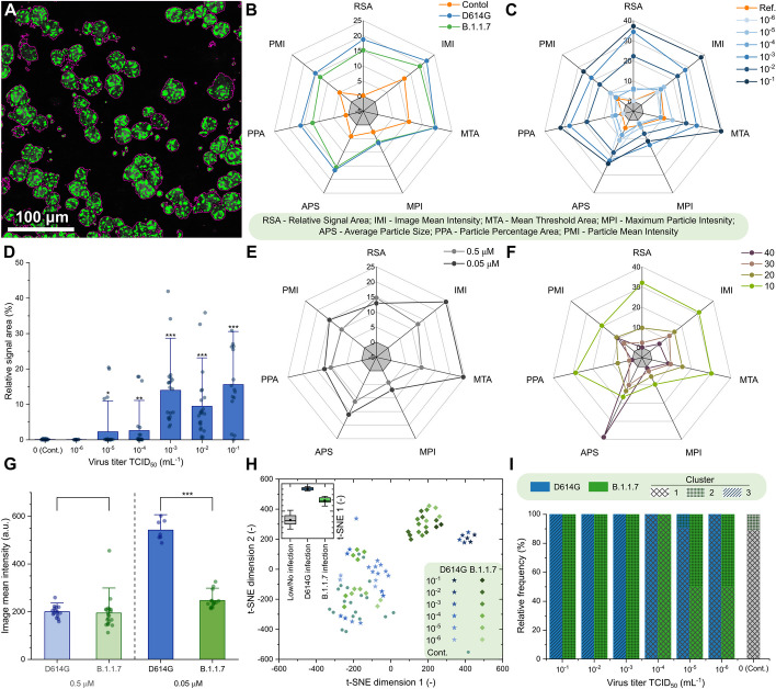
The application of a new calcium sensor molecule is shown, combined with a high-end 2P technique for imaging and identifying the patterns associated with cellular infection damage within cells. Vero E6 cells infected with SARS-CoV-2 variants, D614G or B.1.1.7, exhibit elevated cytosolic calcium levels, allowing infection monitoring by tracking the cellular changes in calcium level by the internalized calcium sensor. The imaging provides valuable information on how the level and intracellular distribution of calcium are perturbed during the infection. Moreover, two-photon calcium sensing allowed the distinction of infections by two studied viral variants via cluster analysis of the image parameters. This approach will facilitate the study of cellular correlates of infection and their quantification depending on viral variants and viral load.
We propose a new two-photon microscopy-based method combined with a cell-internalized sensor to quantify the level of SARS-CoV-2 infection. We optimized the applied dye concentrations to not interfere with viral fusion and viral replication events. The presented method ensured the proper monitoring of viral infection, replication, and cell fate. It also enabled distinguishing intracellular details of cell damage, such as vacuole and apoptotic body formation. Using clustering analysis, 2P microscopy calcium fluorescence images were suitable to distinguish two different viral variants in cell cultures. Cellular harm levels read out by calcium imaging were quantitatively related to the initial viral multiplicity of infection numbers. Thus, 2P quantitative calcium imaging might be used as a correlate of infection or a correlate of activity in cellular antiviral studies.
严重急性呼吸综合征冠状病毒 2 (SARS-CoV-2) 病毒感染对机体的影响已得到广泛研究,但由于缺乏合适的高分辨率实验系统,对于感染如何在细胞间或细胞内随时间传播的动力学知之甚少。据报道,SARS-CoV-2 感染途径集中在钙内流和亚细胞钙分布变化上。成像结合适当的染色技术是研究亚细胞钙相关感染和复制机制的有效工具。
使用双光子(2P)荧光成像和我们新的钙选择性染料,应用自动图像分析和聚类分析来揭示滴度和变体效应对 SARS-CoV-2 感染的 Vero E6 细胞的影响。
展示了一种新的钙传感器分子的应用,结合高端 2P 技术对细胞内与细胞感染损伤相关的模式进行成像和识别。感染 SARS-CoV-2 变体 D614G 或 B.1.1.7 的 Vero E6 细胞显示出细胞溶质钙水平升高,通过跟踪细胞内钙传感器内化引起的钙水平变化,可以对感染进行监测。该成像提供了有关钙水平和细胞内分布在感染过程中如何受到干扰的有价值信息。此外,双光子钙感应通过对图像参数进行聚类分析,能够区分两种研究病毒变体的感染。这种方法将有助于研究感染的细胞相关性及其依赖于病毒变体和病毒载量的量化。
我们提出了一种新的基于双光子显微镜的方法,结合细胞内传感器来定量 SARS-CoV-2 感染。我们优化了应用染料浓度,使其不干扰病毒融合和病毒复制事件。所提出的方法确保了对病毒感染、复制和细胞命运的适当监测。它还能够区分细胞损伤的细胞内细节,如空泡和凋亡小体的形成。使用聚类分析,2P 显微镜钙荧光图像可用于区分细胞培养物中的两种不同病毒变体。钙成像读出的细胞损伤水平与初始病毒感染复数数量定量相关。因此,2P 定量钙成像可用于作为细胞抗病毒研究中的感染相关或活性相关指标。