Otolaryngology / Head and Neck Surgery, Amsterdam UMC Locatie VUmc, Amsterdam, The Netherlands.
Cancer Biology and Immunology, Cancer Centre Amsterdam, Amsterdam, The Netherlands.
J Immunother Cancer. 2024 Jul 24;12(7):e009550. doi: 10.1136/jitc-2024-009550.
Approximately 50% of head and neck squamous cell carcinomas (HNSCC) recur after treatment with curative intent. Immune checkpoint inhibitors are treatment options for recurrent/metastatic HNSCC; however, less than 20% of patients respond. To increase this response rate, it is fundamental to increase our understanding of the spatial tumor immune microenvironment (TIME).
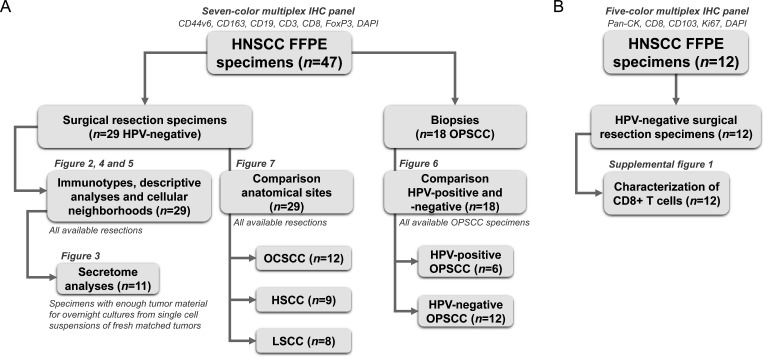
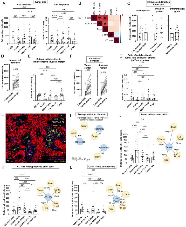
In total, 53 HNSCC specimens were included. Using a seven-color multiplex immunohistochemistry panel we identified tumor cells, CD163+macrophages, B cells, CD8+T cells, CD4+T helper cells and regulatory T cells (Tregs) in treatment-naive surgical resection specimens (n=29) and biopsies (n=18). To further characterize tumor-infiltrating CD8+T cells, we stained surgical resection specimens (n=12) with a five-color tumor-resident panel including CD103, Ki67, CD8 and pan-cytokeratin. Secretome analysis was performed on matched tumor suspensions (n=11) to measure protein levels.
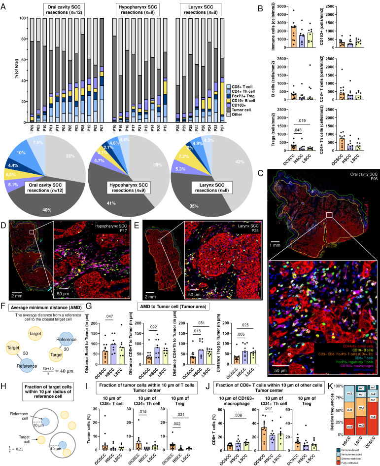
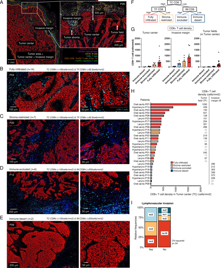
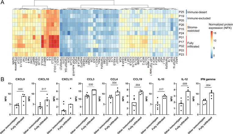
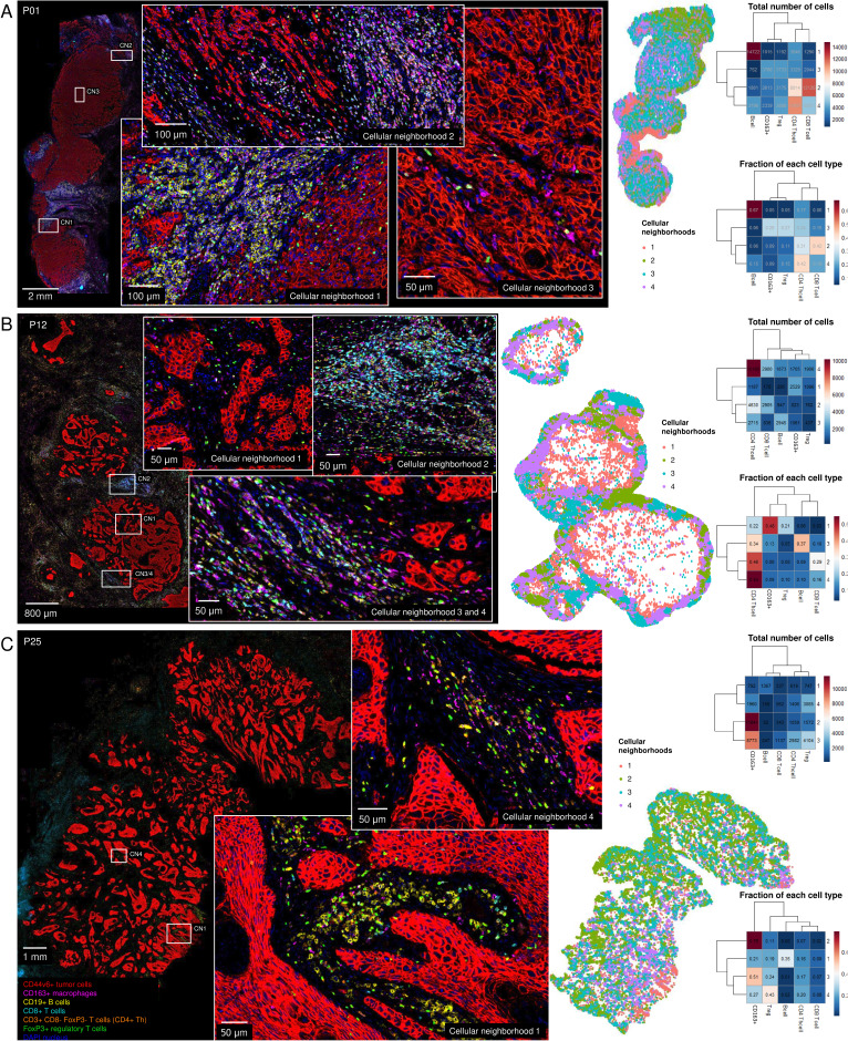
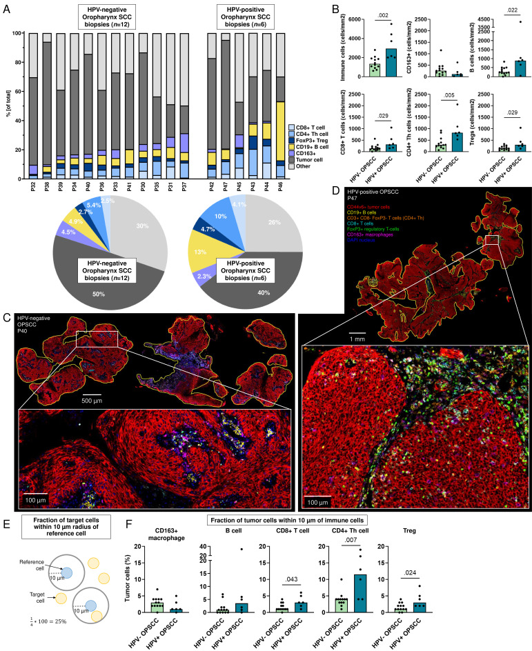
Based on CD8+T cell infiltrates, we identified four different immunotypes: fully infiltrated, stroma-restricted, immune-excluded, and immune-desert. We found higher cytokine levels in fully infiltrated tumors compared with other immunotypes. While the highest immune infiltrates were observed in the invasive margin for all immune cells, CD163+macrophages and Tregs had the highest tendency to infiltrate the tumor center. Within the tumor center, especially B cells stayed at the tumor stroma, whereas CD163+macrophages, followed by T cells, were more often localized within tumor fields. Also, B cells were found further away from other cells and often formed aggregates while T cells and CD163+macrophages tended to be more closely located to each other. Across resection specimens from various anatomical sites within the head and neck, oral cavity tumors exhibited the highest densities of Tregs. Moreover, the distance from B cells and T cells to tumor cells was shortest in oral cavity squamous cell carcinoma (OCSCC), suggesting more interaction between lymphocytes and tumor cells. Also, the fraction of T cells within 10 µm of CD163+macrophages was lowest in OCSCC, indicating fewer myeloid/T-cell suppressive interactions in OCSCC.
We comprehensively described the TIME of HNSCC using a unique data set of resection specimens. We discovered that the composition, as well as the relative localization of immune cells in the TIME, differed in distinct anatomical sites of the head and neck.
约 50%的头颈部鳞状细胞癌(HNSCC)在接受根治性治疗后复发。免疫检查点抑制剂是复发性/转移性 HNSCC 的治疗选择;然而,只有不到 20%的患者有反应。为了提高这种反应率,必须提高我们对空间肿瘤免疫微环境(TIME)的理解。
总共纳入了 53 例 HNSCC 标本。我们使用七色多重免疫组化试剂盒鉴定了治疗前手术切除标本(n=29)和活检(n=18)中的肿瘤细胞、CD163+巨噬细胞、B 细胞、CD8+T 细胞、CD4+辅助性 T 细胞和调节性 T 细胞(Tregs)。为了进一步描述肿瘤浸润性 CD8+T 细胞,我们用包括 CD103、Ki67、CD8 和泛细胞角蛋白在内的五色肿瘤驻留试剂盒对 12 例手术切除标本进行染色。对匹配的肿瘤悬液(n=11)进行分泌组分析以测量蛋白水平。
根据 CD8+T 细胞浸润情况,我们鉴定了四种不同的免疫表型:完全浸润型、基质受限型、免疫排除型和免疫荒漠型。我们发现与其他免疫表型相比,完全浸润型肿瘤的细胞因子水平更高。虽然所有免疫细胞在侵袭边缘的免疫浸润最高,但 CD163+巨噬细胞和 Tregs 有更高的倾向浸润肿瘤中心。在肿瘤中心,B 细胞尤其位于肿瘤基质中,而 CD163+巨噬细胞紧随其后,更常位于肿瘤区域内。此外,B 细胞与其他细胞的距离更远,通常形成聚集体,而 T 细胞和 CD163+巨噬细胞往往彼此更靠近。在头颈部不同解剖部位的切除标本中,口腔鳞状细胞癌(OCSCC)的 Tregs 密度最高。此外,在口腔鳞状细胞癌中,B 细胞和 T 细胞与肿瘤细胞的距离最短,提示淋巴细胞与肿瘤细胞之间的相互作用更多。而且,在 OCSCC 中,CD163+巨噬细胞 10μm 内的 T 细胞比例最低,表明 OCSCC 中骨髓/T 细胞抑制性相互作用较少。
我们使用独特的切除标本数据集全面描述了 HNSCC 的 TIME。我们发现,在不同的头颈部解剖部位,TIME 中免疫细胞的组成以及相对定位存在差异。