Litwin Tomasz, Rędzia-Ogrodnik Barbara, Antos Agnieszka, Przybyłkowski Adam, Członkowska Anna, Bembenek Jan Paweł
Second Department of Neurology, Institute of Psychiatry and Neurology, 02-957 Warsaw, Poland.
Department of Gastroenterology, Medical University, Warsaw 02-097, Poland.
Brain Sci. 2024 Jul 19;14(7):727. doi: 10.3390/brainsci14070727.
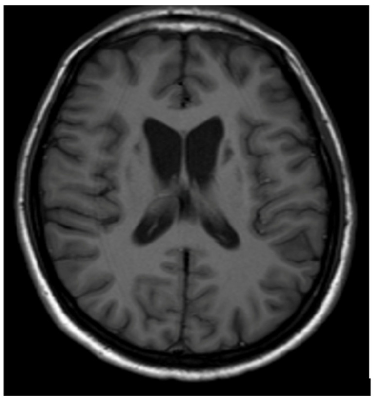
Wilson's disease (WD) is a genetic disorder of copper metabolism with pathological copper accumulation in many organs, resulting in clinical symptoms, mostly hepatic and neuropsychiatric. As copper accumulates in the brain during WD, and almost 50% of WD patients at diagnosis present with neurological symptoms, neuroimaging studies (especially brain magnetic resonance imaging (MRI)) are part of WD diagnosis. The classical sequences (T1, T2, and fluid-attenuated inversion recovery) were used to describe brain MRI; however, with the development of neuroradiology, several papers proposed the use of new MRI sequences and techniques like susceptibility-weighted images, T2*, diffusion MRI, tractography, volumetric assessment and post-processing brain MRI analysis of paramagnetic accumulation-quantitative susceptibility mapping. Based on these neuroradiological data in WD, currently, brain MRI semiquantitative scale and the pathognomonic neuroradiological brain MRI signs in WD were proposed. Further, the volumetric studies and brain iron accumulation MRI analysis suggested brain atrophy and iron accumulation as biomarkers of neurological WD disease severity. All these results highlight the significance of brain MRI examinations in WD. Due to the extreme progress of these studies, based on the available literature, the authors present the current state of knowledge about the significance, practical aspects, and future directions of brain MRI in WD.
威尔逊病(WD)是一种铜代谢的遗传性疾病,病理状态下铜在许多器官中蓄积,导致临床症状,主要为肝脏和神经精神方面的症状。由于在WD病程中铜在脑内蓄积,且几乎50%的WD患者在诊断时出现神经症状,神经影像学检查(尤其是脑磁共振成像(MRI))是WD诊断的一部分。经典序列(T1、T2和液体衰减反转恢复序列)曾用于描述脑部MRI;然而,随着神经放射学的发展,一些论文提出使用新的MRI序列和技术,如磁敏感加权成像、T2*、扩散MRI、纤维束成像、容积评估以及对顺磁性物质蓄积进行后处理的脑MRI分析——定量磁敏感成像。基于WD的这些神经放射学数据,目前提出了脑MRI半定量量表以及WD具有诊断意义的神经放射学脑MRI征象。此外,容积研究和脑铁蓄积MRI分析提示脑萎缩和铁蓄积可作为神经型WD疾病严重程度的生物标志物。所有这些结果都凸显了脑MRI检查在WD中的重要性。鉴于这些研究取得的巨大进展,基于现有文献,作者阐述了关于脑MRI在WD中的意义、实际应用及未来方向的当前知识状况。