• 文献检索
  • 文档翻译
  • 深度研究
  • 学术资讯
  • Suppr Zotero 插件Zotero 插件
  • 邀请有礼
  • 套餐&价格
  • 历史记录
应用&插件
Suppr Zotero 插件Zotero 插件浏览器插件Mac 客户端Windows 客户端微信小程序
定价
高级版会员购买积分包购买API积分包
服务
文献检索文档翻译深度研究API 文档MCP 服务
关于我们
关于 Suppr公司介绍联系我们用户协议隐私条款
关注我们

Suppr 超能文献

核心技术专利:CN118964589B侵权必究
粤ICP备2023148730 号-1Suppr @ 2026

文献检索

告别复杂PubMed语法,用中文像聊天一样搜索,搜遍4000万医学文献。AI智能推荐,让科研检索更轻松。

立即免费搜索

文件翻译

保留排版,准确专业,支持PDF/Word/PPT等文件格式,支持 12+语言互译。

免费翻译文档

深度研究

AI帮你快速写综述,25分钟生成高质量综述,智能提取关键信息,辅助科研写作。

立即免费体验

缺氧诱导因子1α通过调控自噬影响牦牛卵母细胞成熟和早期胚胎发育。

Hypoxia-Inducible Factor 1α Affects Yak Oocyte Maturation and Early Embryonic Development by Regulating Autophagy.

作者信息

Ma Xin, Wang Meng, Wang Jinglei, Han Xiaohong, Yang Xiaoqing, Zhang Hui, Zhong Donglan, Qiu Shantong, Yu Sijiu, Wang Libin, Pan Yangyang

机构信息

College of Veterinary Medicine, Gansu Agricultural University, Lanzhou 730070, China.

Gansu Province Livestock Embryo Engineering Research Center, Lanzhou 730070, China.

出版信息

Antioxidants (Basel). 2024 Jul 14;13(7):840. doi: 10.3390/antiox13070840.

DOI:10.3390/antiox13070840
PMID:39061908
原文链接:https://pmc.ncbi.nlm.nih.gov/articles/PMC11273763/
Abstract

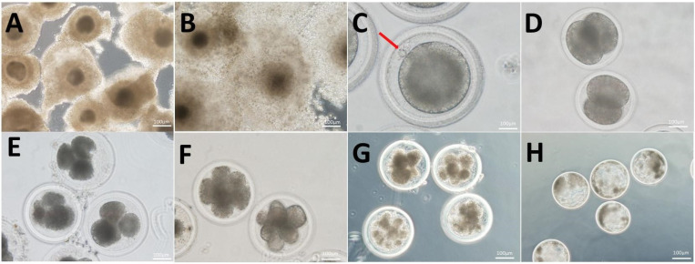
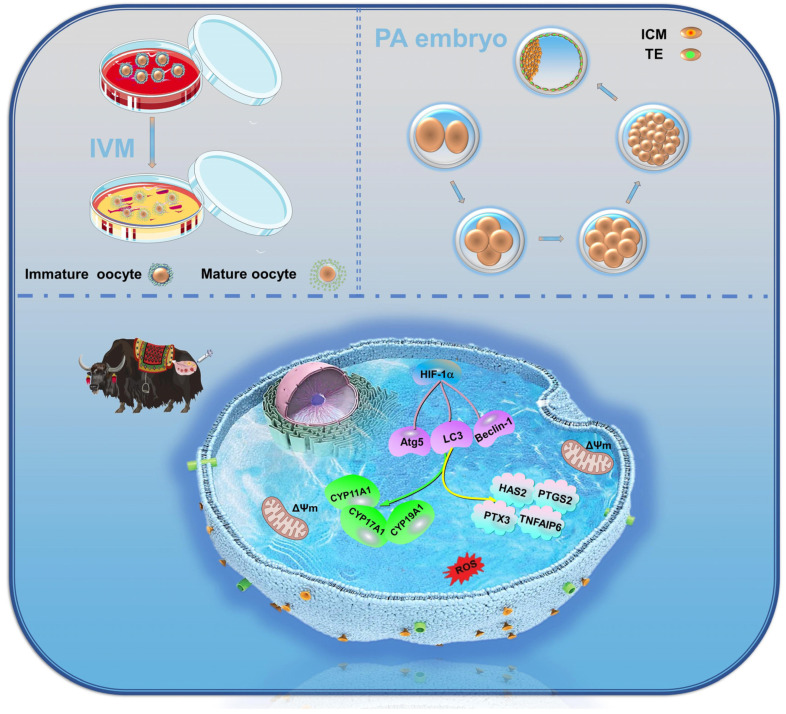
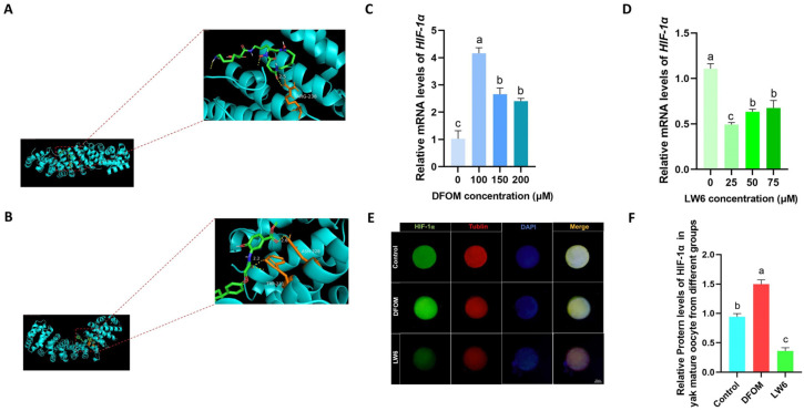
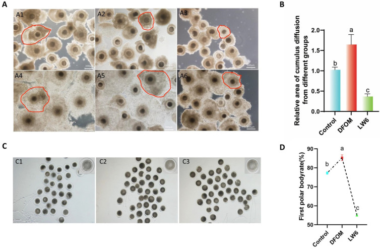
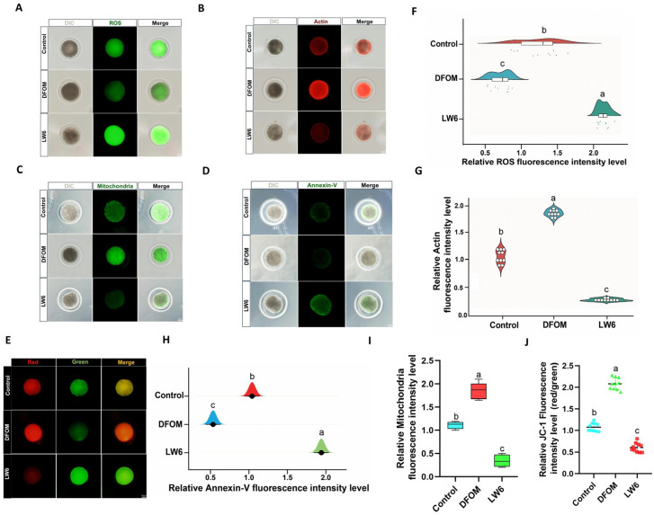
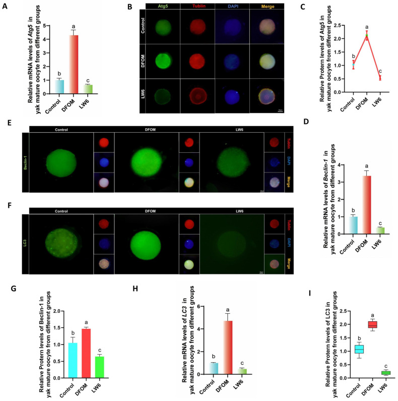
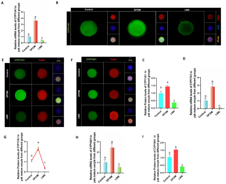
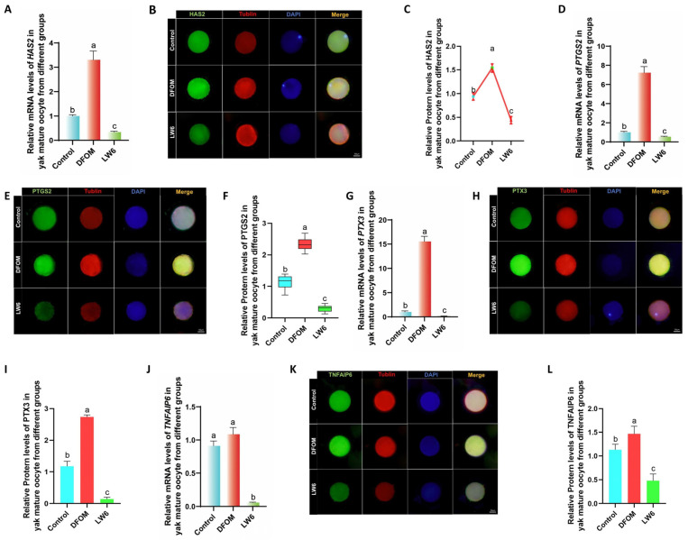
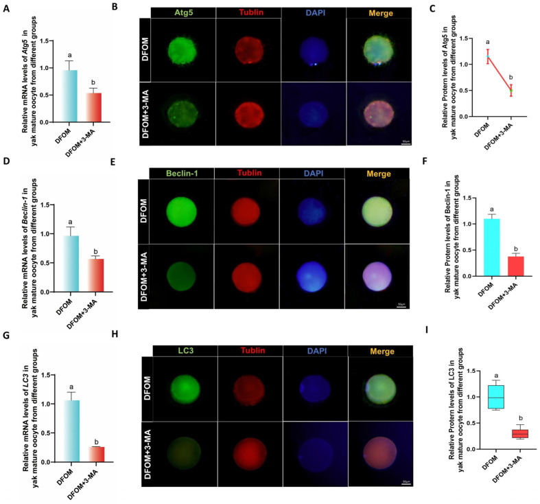
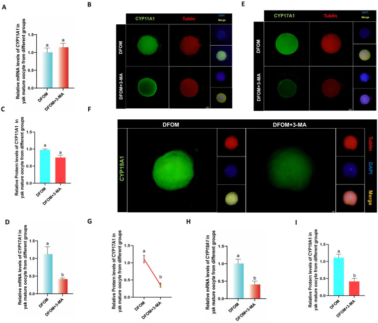
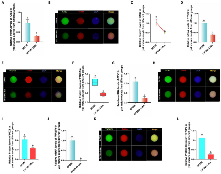
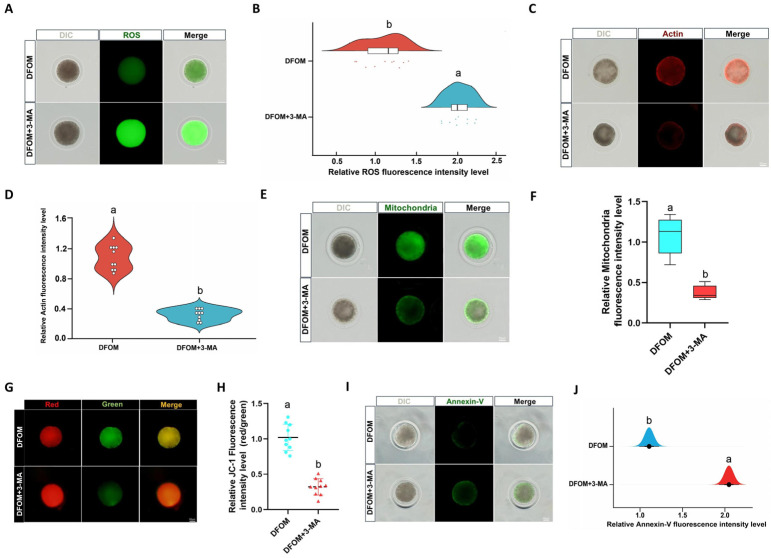
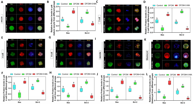
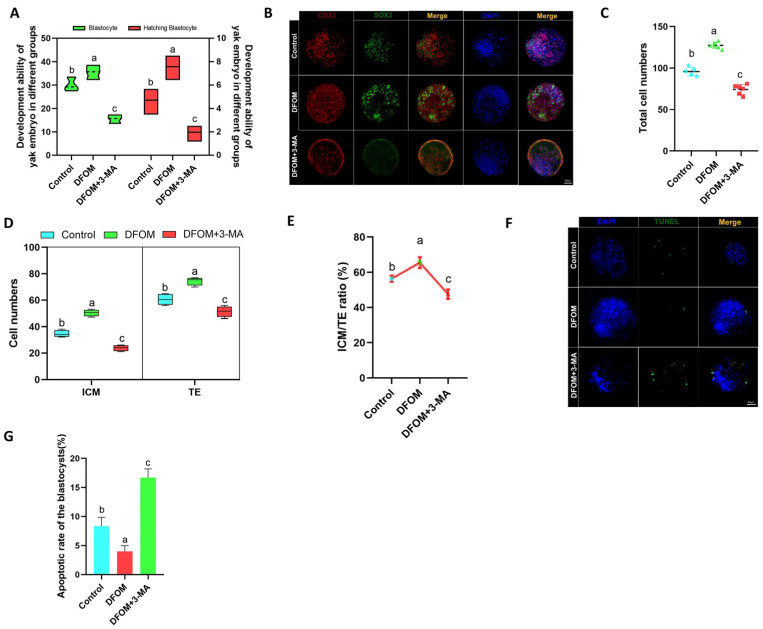
In animal assisted reproductive technology, the production of high-quality oocytes is crucial. The yak, having lived in the Qinghai-Tibet Plateau for an extended period, has reproductive cells that are regulated by hypoxia-inducible factor 1α (HIF-1α). This study aimed to investigate the impact of HIF-1α on yak oocyte maturation and early embryonic development in vitro through the regulation of autophagy. The in vitro maturation process of yak oocytes involved the addition of the HIF-1α inducer DFOM and the inhibitor LW6 to examine their effects on yak oocyte maturation, early embryonic development, cell autophagy, cytochrome P450s (CYP450s) enzyme expression, and cumulus diffusion factors. The findings revealed that DFOM significantly upregulated the expression of HIF-1α, resulting in increased the cumulus diffusion area, elevated first polar body expulsion rate of oocytes, enhanced mitochondrial and actin levels, decreased ROS production, and reduced early apoptosis levels of oocytes. Moreover, DFOM promoted the expression of autophagy-related proteins, CYP450s enzymes, and cumulus diffusion factors, thereby enhancing oocyte maturation and early embryonic development. Conversely, LW6 exhibited opposite effects. The inhibition of autophagy levels with 3-MA during DFOM treatment yielded similar outcomes. Furthermore, reducing autophagy led to increased apoptosis levels at all stages of early embryonic development, as well as a significant decrease in total cell number and ICM/TE ratio of blastocysts. Studies have shown that during the in vitro maturation of yak oocytes, HIF-1α can affect the cumulus expansion area of oocytes by regulating autophagy, the first polar body excretion rate, mitochondrial level, actin level, ROS and early apoptosis level, the CYP450s enzyme, and the expression of cumulus expansion factors, thereby improving the in vitro maturation and early embryonic development of yak oocytes. These findings offer valuable insights into the reproductive regulation mechanism of yaks in hypoxic environments and suggest potential strategies for the advancement of yak assisted reproductive technology.

摘要

在动物辅助生殖技术中,高质量卵母细胞的产生至关重要。牦牛长期生活在青藏高原,其生殖细胞受缺氧诱导因子1α(HIF-1α)调控。本研究旨在通过自噬调节来探究HIF-1α对牦牛卵母细胞体外成熟和早期胚胎发育的影响。牦牛卵母细胞的体外成熟过程中添加了HIF-1α诱导剂DFOM和抑制剂LW6,以检测它们对牦牛卵母细胞成熟、早期胚胎发育、细胞自噬、细胞色素P450s(CYP450s)酶表达和卵丘扩散因子的影响。研究结果表明,DFOM显著上调HIF-1α的表达,导致卵丘扩散面积增加、卵母细胞第一极体排出率升高、线粒体和肌动蛋白水平提高、活性氧产生减少以及卵母细胞早期凋亡水平降低。此外,DFOM促进自噬相关蛋白、CYP450s酶和卵丘扩散因子的表达,从而增强卵母细胞成熟和早期胚胎发育。相反,LW6表现出相反的效果。在DFOM处理期间用3-MA抑制自噬水平产生了类似的结果。此外,减少自噬导致早期胚胎发育各阶段的凋亡水平增加,以及囊胚的总细胞数和ICM/TE比率显著降低。研究表明,在牦牛卵母细胞体外成熟过程中,HIF-1α可通过调节自噬影响卵母细胞的卵丘扩展面积、第一极体排出率、线粒体水平、肌动蛋白水平、活性氧和早期凋亡水平、CYP450s酶以及卵丘扩展因子的表达,从而改善牦牛卵母细胞的体外成熟和早期胚胎发育。这些发现为牦牛在缺氧环境中的生殖调控机制提供了有价值的见解,并为牦牛辅助生殖技术的进步提出了潜在策略。

https://cdn.ncbi.nlm.nih.gov/pmc/blobs/073f/11273763/f868efcb0a00/antioxidants-13-00840-g014.jpg
https://cdn.ncbi.nlm.nih.gov/pmc/blobs/073f/11273763/eaa63fa5b820/antioxidants-13-00840-g001.jpg
https://cdn.ncbi.nlm.nih.gov/pmc/blobs/073f/11273763/9ec310807dc5/antioxidants-13-00840-g002.jpg
https://cdn.ncbi.nlm.nih.gov/pmc/blobs/073f/11273763/b95307a3f7b8/antioxidants-13-00840-g003.jpg
https://cdn.ncbi.nlm.nih.gov/pmc/blobs/073f/11273763/dd38e6f52137/antioxidants-13-00840-g004.jpg
https://cdn.ncbi.nlm.nih.gov/pmc/blobs/073f/11273763/8040312dd43e/antioxidants-13-00840-g005.jpg
https://cdn.ncbi.nlm.nih.gov/pmc/blobs/073f/11273763/cf1e82c4f917/antioxidants-13-00840-g006.jpg
https://cdn.ncbi.nlm.nih.gov/pmc/blobs/073f/11273763/af2d6142bf03/antioxidants-13-00840-g007.jpg
https://cdn.ncbi.nlm.nih.gov/pmc/blobs/073f/11273763/a11d4a1fba07/antioxidants-13-00840-g008.jpg
https://cdn.ncbi.nlm.nih.gov/pmc/blobs/073f/11273763/b12a89122501/antioxidants-13-00840-g009.jpg
https://cdn.ncbi.nlm.nih.gov/pmc/blobs/073f/11273763/cb5c1ea7ad6c/antioxidants-13-00840-g010.jpg
https://cdn.ncbi.nlm.nih.gov/pmc/blobs/073f/11273763/242912271062/antioxidants-13-00840-g011.jpg
https://cdn.ncbi.nlm.nih.gov/pmc/blobs/073f/11273763/8d426eac13a1/antioxidants-13-00840-g012.jpg
https://cdn.ncbi.nlm.nih.gov/pmc/blobs/073f/11273763/847437f72ead/antioxidants-13-00840-g013.jpg
https://cdn.ncbi.nlm.nih.gov/pmc/blobs/073f/11273763/f868efcb0a00/antioxidants-13-00840-g014.jpg

相似文献

1
Hypoxia-Inducible Factor 1α Affects Yak Oocyte Maturation and Early Embryonic Development by Regulating Autophagy.缺氧诱导因子1α通过调控自噬影响牦牛卵母细胞成熟和早期胚胎发育。
Antioxidants (Basel). 2024 Jul 14;13(7):840. doi: 10.3390/antiox13070840.
2
Low oxygen concentration improves yak oocyte maturation and inhibits apoptosis through HIF-1 and VEGF.低氧浓度通过 HIF-1 和 VEGF 提高牦牛卵母细胞成熟并抑制凋亡。
Reprod Domest Anim. 2022 Apr;57(4):381-392. doi: 10.1111/rda.14076. Epub 2022 Jan 21.
3
Effect of rapamycin treatment on oocyte in vitro maturation and embryonic development after parthenogenesis in yaks.雷帕霉素处理对牦牛孤雌生殖后卵母细胞体外成熟和胚胎发育的影响。
Theriogenology. 2022 Nov;193:128-135. doi: 10.1016/j.theriogenology.2022.09.017. Epub 2022 Sep 18.
4
Expression patterns of ZO-1/2 and their effects on porcine oocyte in vitro maturation and early embryonic development.紧密连接蛋白1/2(ZO-1/2)的表达模式及其对猪卵母细胞体外成熟和早期胚胎发育的影响
Theriogenology. 2021 Feb;161:262-270. doi: 10.1016/j.theriogenology.2020.12.009. Epub 2020 Dec 13.
5
Low oxygen concentrations improve yak oocyte maturation and enhance the developmental competence of preimplantation embryos.低氧浓度可改善牦牛卵母细胞成熟并增强植入前胚胎的发育能力。
Theriogenology. 2020 Oct 15;156:46-58. doi: 10.1016/j.theriogenology.2020.06.022. Epub 2020 Jun 28.
6
Follistatin (FST) is expressed in buffalo (Bubalus bubalis) ovarian follicles and promotes oocyte maturation and early embryonic development.卵泡抑素 (Follistatin, FST) 在水牛 (Bubalus bubalis) 卵巢滤泡中表达,可促进卵母细胞成熟和早期胚胎发育。
Reprod Domest Anim. 2023 Dec;58(12):1718-1731. doi: 10.1111/rda.14490. Epub 2023 Nov 2.
7
Regulatory effects of HIF-1α and HO-1 in hypoxia-induced proliferation of pulmonary arterial smooth muscle cells in yak.低氧诱导牦牛肺动脉平滑肌细胞增殖中HIF-1α和HO-1的调控作用
Cell Signal. 2021 Nov;87:110140. doi: 10.1016/j.cellsig.2021.110140. Epub 2021 Aug 31.
8
Leukemia inhibitory factor enhances the development and subsequent blastocysts quality of yak oocytes .白血病抑制因子可提高牦牛卵母细胞的发育能力及随后的囊胚质量。
Front Vet Sci. 2022 Sep 21;9:997709. doi: 10.3389/fvets.2022.997709. eCollection 2022.
9
Treatment with acetyl-l-carnitine during in vitro maturation of buffalo oocytes improves oocyte quality and subsequent embryonic development.在水牛卵母细胞体外成熟过程中用乙酰左旋肉碱处理可提高卵母细胞质量及后续胚胎发育。
Theriogenology. 2018 Sep 15;118:80-89. doi: 10.1016/j.theriogenology.2018.05.033. Epub 2018 Jun 1.
10
Glycine regulates lipid peroxidation promoting porcine oocyte maturation and early embryonic development.甘氨酸调节脂质过氧化,促进猪卵母细胞成熟和早期胚胎发育。
J Anim Sci. 2023 Jan 3;101. doi: 10.1093/jas/skac425.

引用本文的文献

1
Gut Microbiota and Its Metabolites Modulate Pregnancy Outcomes by Regulating Placental Autophagy and Ferroptosis.肠道微生物群及其代谢产物通过调节胎盘自噬和铁死亡来调节妊娠结局。
Antioxidants (Basel). 2025 Aug 7;14(8):970. doi: 10.3390/antiox14080970.
2
CIRBP Enhances the Function of Yak Cumulus Cells by Activating AMPK/mTOR-Mediated Mitophagy.CIRBP通过激活AMPK/mTOR介导的线粒体自噬增强牦牛卵丘细胞的功能。
Biomolecules. 2025 May 24;15(6):759. doi: 10.3390/biom15060759.
3
Cloning, bioinformatics analysis and expression of the cysteine dioxygenase type 1 (CDO1) gene in domestic yak.

本文引用的文献

1
Hypoxia enhances autophagy level of human sperms.缺氧可增强人精子的自噬水平。
Sci Rep. 2024 Apr 11;14(1):8465. doi: 10.1038/s41598-024-59213-1.
2
Unravelling the role of HAS2, GREM1, and PTGS2 gene expression in cumulus cells: implications for human oocyte development competency - a systematic review and integrated bioinformatic analysis.解开 HAS2、GREM1 和 PTGS2 基因表达在卵丘细胞中的作用:对人类卵母细胞发育能力的影响——系统评价和综合生物信息学分析。
Front Endocrinol (Lausanne). 2024 Mar 8;15:1274376. doi: 10.3389/fendo.2024.1274376. eCollection 2024.
3
Mapping conservation priorities for wild yak (Bos mutus) habitats on the Tibetan Plateau, China.
家牦牛中1型半胱氨酸双加氧酶(CDO1)基因的克隆、生物信息学分析及表达
Front Vet Sci. 2024 Oct 18;11:1488782. doi: 10.3389/fvets.2024.1488782. eCollection 2024.
中国青藏高原野生牦牛(Bos mutus)栖息地保护重点的划定。
Sci Total Environ. 2024 Mar 1;914:169803. doi: 10.1016/j.scitotenv.2023.169803. Epub 2024 Jan 4.
4
Expression and regulation of HIF-1a in hypoxic pulmonary hypertension: Focus on pathological mechanism and Pharmacological Treatment.低氧性肺动脉高压中 HIF-1a 的表达与调控:关注病理机制与药物治疗。
Int J Med Sci. 2024 Jan 1;21(1):45-60. doi: 10.7150/ijms.88216. eCollection 2024.
5
Factors Influencing the Maturation and Developmental Competence of Yak () Oocytes In Vitro.影响牦牛卵母细胞体外成熟和发育能力的因素。
Genes (Basel). 2023 Sep 27;14(10):1882. doi: 10.3390/genes14101882.
6
HIF-1α signaling: Essential roles in tumorigenesis and implications in targeted therapies.缺氧诱导因子-1α信号传导:在肿瘤发生中的关键作用及在靶向治疗中的意义。
Genes Dis. 2023 Mar 30;11(1):234-251. doi: 10.1016/j.gendis.2023.02.039. eCollection 2024 Jan.
7
FOXG1 is involved in mouse ovarian functions and embryogenesis.FOXG1 参与了老鼠的卵巢功能和胚胎发生。
J Steroid Biochem Mol Biol. 2023 Oct;233:106372. doi: 10.1016/j.jsbmb.2023.106372. Epub 2023 Aug 2.
8
Astaxanthin protects oocyte maturation against cypermethrin-induced defects in pigs.虾青素可保护卵母细胞成熟,防止氯氰菊酯对猪造成的缺陷。
Theriogenology. 2023 Oct 1;209:31-39. doi: 10.1016/j.theriogenology.2023.06.022. Epub 2023 Jun 15.
9
Meiosis: Actin and microtubule networks drive chromosome clustering in oocytes.减数分裂:动粒和微管网络驱动卵母细胞中的染色体聚集。
Curr Biol. 2023 Apr 10;33(7):R272-R274. doi: 10.1016/j.cub.2023.02.061.
10
Role of diacerein on steroidogenesis and folliculogenesis related genes in ovary of letrozole-induced PCOS mice.二苯乙烯苷对来曲唑诱导的多囊卵巢综合征小鼠卵巢甾体激素生成和卵泡发生相关基因的作用。
Chem Biol Interact. 2023 May 25;377:110468. doi: 10.1016/j.cbi.2023.110468. Epub 2023 Apr 6.