Fiedler Lukas S, Lippert Burkard M, Adrian Lukas, Meyer Tobias
Department for Otorhinolaryngology/Head and Neck, Plastic Surgery, SLK Kliniken Heilbronn, 74078 Heilbronn, Germany.
Faculty of Medicine, Heidelberg University, 69120 Heidelberg, Germany.
J Pers Med. 2024 Jul 5;14(7):730. doi: 10.3390/jpm14070730.
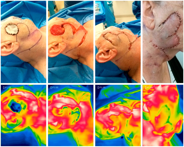
Successful outcomes in head and neck surgery rely on maintaining perfusion in pedicled skin flaps. Thermal imaging offers a noninvasive means to assess tissue perfusion, potentially aiding in predicting flap viability. This pilot study explores the utility of SBTI (smartphone-based thermal imaging) for predicting flap vitality and monitoring during surgery.
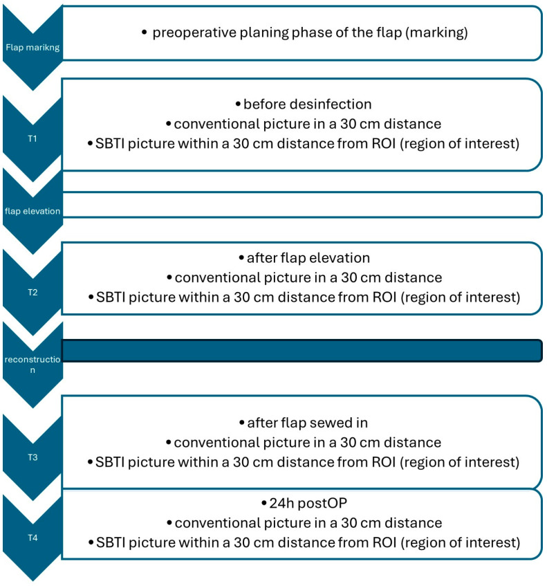
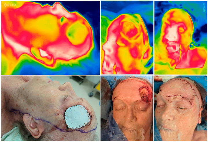
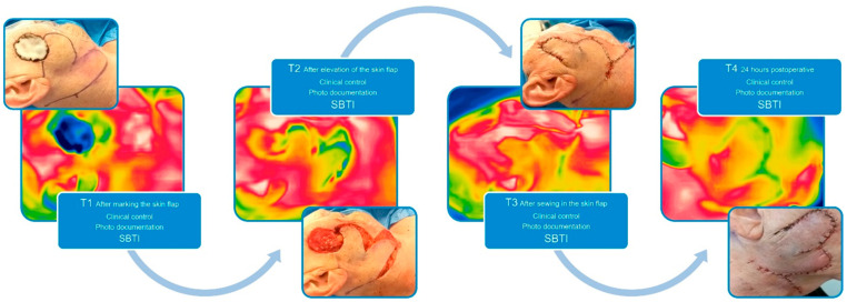
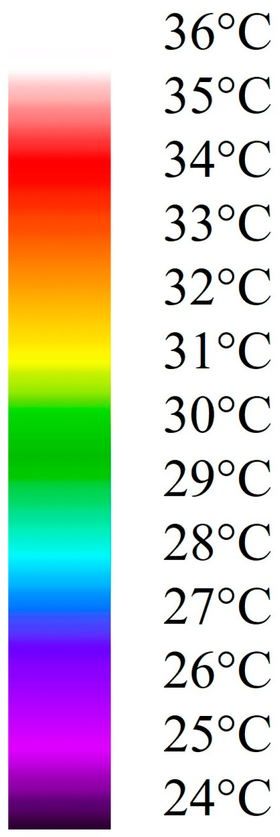
Thermal imaging was employed using the FLIR One System. An imaging protocol was established, defining points of interest (T1-T4) on pedicled skin flaps. Conducted over four months, the study integrated SBTI into reconstructive surgery for the face, head and neck defects post-tumor resections. SBTI's effectiveness was assessed with n = 11 pedicled flaps, capturing images at key stages and correlating them with clinical flap assessment. Thermal images were retrospectively graded by two surgeons, evaluating flap perfusion on a scale from 1 to 5, based on temperature differences (1 = ΔT < 2 °C, 2 = ΔT ≥ 2 °C, 3 = ΔT ≥ 4 °C, 4 = ΔT ≥ 6 °C, and 5 = ΔT ≥ 8 °C), with assessments averaged for consensus and compared with the clinical assessment control group.
The study encountered challenges during implementation, leading to the exclusion of six patients. Patient data included 11 cases with n = 44 SBTI images. Intraoperative assessments consistently showed good perfusion. One postoperative dehiscence was noted, which retrospectively coincided with intraoperative SBTI grading, but not with clinical assessment. Statistical analysis indicated consistent outcomes following clinical and SBTI assessments. Thermal imaging accurately predicted flap viability, although it had limitations with small flaps.
SBTI proved effective, inexpensive, and noninvasive for assessing tissue perfusion, showing promise for predicting flap viability and intraoperative monitoring in head and neck surgery.
头颈外科手术的成功结果依赖于维持带蒂皮瓣的灌注。热成像提供了一种非侵入性手段来评估组织灌注,可能有助于预测皮瓣的存活情况。这项初步研究探讨了基于智能手机的热成像(SBTI)在预测皮瓣活力及手术过程中监测的效用。
使用FLIR One系统进行热成像。建立了成像方案,确定带蒂皮瓣上的感兴趣点(T1 - T4)。该研究历时四个月,将SBTI纳入面部、头部和颈部肿瘤切除术后缺损的重建手术中。对11个带蒂皮瓣评估SBTI的有效性,在关键阶段采集图像并将其与临床皮瓣评估相关联。两名外科医生对热图像进行回顾性分级,根据温度差异(1 = ΔT < 2°C,2 = ΔT ≥ 2°C,3 = ΔT ≥ 4°C,4 = ΔT ≥ 6°C以及5 = ΔT ≥ 8°C)将皮瓣灌注评估为1至5级,评估结果取平均值以达成共识,并与临床评估对照组进行比较。
研究在实施过程中遇到挑战,导致6名患者被排除。患者数据包括11例共44张SBTI图像。术中评估始终显示灌注良好。记录到1例术后裂开,回顾性分析发现这与术中SBTI分级相符,但与临床评估不符。统计分析表明临床和SBTI评估结果一致。热成像准确地预测了皮瓣的存活情况,尽管对于小皮瓣存在局限性。
SBTI被证明在评估组织灌注方面有效、廉价且非侵入性,对头颈外科手术中预测皮瓣活力和术中监测具有前景。