• 文献检索
  • 文档翻译
  • 深度研究
  • 学术资讯
  • Suppr Zotero 插件Zotero 插件
  • 邀请有礼
  • 套餐&价格
  • 历史记录
应用&插件
Suppr Zotero 插件Zotero 插件浏览器插件Mac 客户端Windows 客户端微信小程序
定价
高级版会员购买积分包购买API积分包
服务
文献检索文档翻译深度研究API 文档MCP 服务
关于我们
关于 Suppr公司介绍联系我们用户协议隐私条款
关注我们

Suppr 超能文献

核心技术专利:CN118964589B侵权必究
粤ICP备2023148730 号-1Suppr @ 2026

文献检索

告别复杂PubMed语法,用中文像聊天一样搜索,搜遍4000万医学文献。AI智能推荐,让科研检索更轻松。

立即免费搜索

文件翻译

保留排版,准确专业,支持PDF/Word/PPT等文件格式,支持 12+语言互译。

免费翻译文档

深度研究

AI帮你快速写综述,25分钟生成高质量综述,智能提取关键信息,辅助科研写作。

立即免费体验

在肝细胞癌超声筛查中添加造影剂。

Addition of contrast in ultrasound screening for hepatocellular carcinoma.

作者信息

McGillen Kathryn, Aljabban Nabeal, Wu Robert, Shin Benjamin, Schreibman Ian, Luke Franklin, Birkholz James

机构信息

Penn State Health Milton S Hershey Medical Center, Department of Radiology, 500 University Drive, Hershey, PA 17033, USA.

Penn State Health Milton S Hershey Medical Center, Department of Medicine, 500 University Drive, Hershey, PA 17033, USA.

出版信息

Res Diagn Interv Imaging. 2024 Feb 7;9:100039. doi: 10.1016/j.redii.2023.100039. eCollection 2024 Mar.

DOI:10.1016/j.redii.2023.100039
PMID:39076583
原文链接:https://pmc.ncbi.nlm.nih.gov/articles/PMC11265193/
Abstract

OBJECTIVE

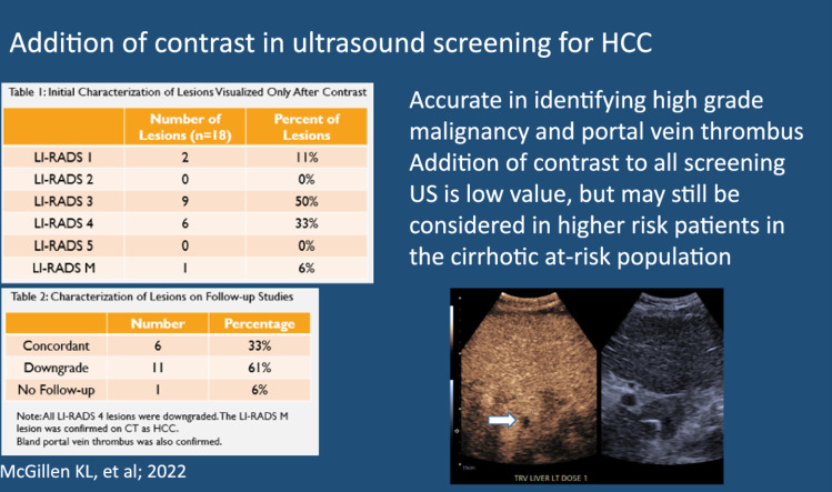
Screening ultrasound for hepatocellular carcinoma (HCC) identifies lesions which require further characterization by a contrast-enhanced exam to non-invasively diagnose HCC. While ultrasound is recommended in screening, some HCC can be occult on grayscale imaging. The purpose of this study was to determine if the addition of ultrasound contrast (sulfahexafluoride) to screening ultrasound for HCC can identify more HCC lesions than grayscale sonographic imaging alone.

METHODS

All HCC screening ultrasounds that also had contrast were evaluated in this retrospective study. Patients with a focal lesion seen only after administration of contrast (OAC) were noted, as well as any follow-up imaging or pathology results. Additional variables collected included patient demographics, cirrhosis type, and laboratory values.

RESULTS

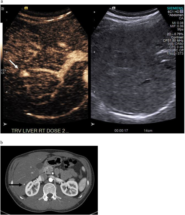
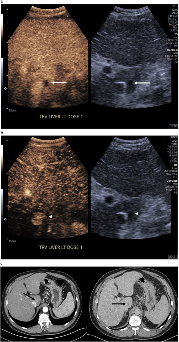
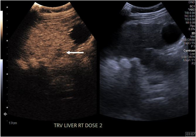
230 unique patients were included, of which 160 had imaging or pathology follow-up. 18 of these patients had an OAC lesion, of which 17 had follow-up. Among these OACs, there was one LIRADS M lesion (1/18, 5.6 %) and one bland portal vein thrombus identified, which were both confirmed on follow-up imaging. All LIRADS 4 OAC lesions were downgraded. No additional HCC were identified on follow-up imaging or pathology of these patients.

CONCLUSION

Addition of contrast to screening ultrasound did identify additional lesions, portal vein thrombus, and high grade malignancy. However, as the incidence of OAC lesions was low (7.8 %, 18/230) and most of the lesions were not malignant, addition of post contrast sweeps through the liver is of low value in the low to medium at-risk cirrhotic population in identifying occult HCC.

摘要

目的

用于肝细胞癌(HCC)筛查的超声检查可发现需要通过增强检查进一步定性以无创诊断HCC的病变。虽然推荐超声用于筛查,但某些HCC在灰阶成像上可能隐匿。本研究的目的是确定在HCC筛查超声中添加超声造影剂(六氟化硫)是否比单纯灰阶超声成像能发现更多的HCC病变。

方法

在这项回顾性研究中评估了所有同时进行了超声造影的HCC筛查超声检查。记录仅在注射造影剂后才发现局灶性病变(OAC)的患者,以及任何后续的成像或病理结果。收集的其他变量包括患者人口统计学特征、肝硬化类型和实验室值。

结果

纳入了230例独特患者,其中160例有成像或病理随访。这些患者中有18例有OAC病变,其中17例有随访。在这些OAC病变中,有1例LIRADS M类病变(1/18,5.6%)和1例单纯门静脉血栓被发现,两者均在后续成像中得到证实。所有LIRADS 4类OAC病变均被降级。在这些患者的后续成像或病理检查中未发现其他HCC。

结论

在筛查超声中添加造影剂确实发现了额外的病变、门静脉血栓和高级别恶性肿瘤。然而,由于OAC病变的发生率较低(7.8%,18/230)且大多数病变并非恶性,在中低风险肝硬化人群中,对肝脏进行造影剂增强扫描在识别隐匿性HCC方面价值较低。

https://cdn.ncbi.nlm.nih.gov/pmc/blobs/d6d0/11265193/1ac0237a2ac6/gr3.jpg
https://cdn.ncbi.nlm.nih.gov/pmc/blobs/d6d0/11265193/a2ac4da05225/ga1.jpg
https://cdn.ncbi.nlm.nih.gov/pmc/blobs/d6d0/11265193/48a13c099b4f/gr1.jpg
https://cdn.ncbi.nlm.nih.gov/pmc/blobs/d6d0/11265193/c95df5820e0b/gr2.jpg
https://cdn.ncbi.nlm.nih.gov/pmc/blobs/d6d0/11265193/1ac0237a2ac6/gr3.jpg
https://cdn.ncbi.nlm.nih.gov/pmc/blobs/d6d0/11265193/a2ac4da05225/ga1.jpg
https://cdn.ncbi.nlm.nih.gov/pmc/blobs/d6d0/11265193/48a13c099b4f/gr1.jpg
https://cdn.ncbi.nlm.nih.gov/pmc/blobs/d6d0/11265193/c95df5820e0b/gr2.jpg
https://cdn.ncbi.nlm.nih.gov/pmc/blobs/d6d0/11265193/1ac0237a2ac6/gr3.jpg

相似文献

1
Addition of contrast in ultrasound screening for hepatocellular carcinoma.在肝细胞癌超声筛查中添加造影剂。
Res Diagn Interv Imaging. 2024 Feb 7;9:100039. doi: 10.1016/j.redii.2023.100039. eCollection 2024 Mar.
2
B-mode and contrast enhanced ultrasound guided biopsy of portal vein thrombosis. Value in the diagnosis of occult hepatocellular carcinoma in liver cirrhosis.B 型及超声造影引导下门静脉血栓活检。对肝硬化隐匿性肝细胞癌的诊断价值。
Med Ultrason. 2010 Dec;12(4):286-94.
3
Contrast-enhanced ultrasound for screening hepatocellular carcinoma: an implemented program at a semi-rural academic center.超声造影在肝细胞癌筛查中的应用:半农村地区学术中心的实施项目。
Abdom Radiol (NY). 2021 Sep;46(9):4170-4177. doi: 10.1007/s00261-021-03104-w. Epub 2021 May 6.
4
Imaging Features of Hepatocellular Carcinoma in the Non-Cirrhotic Liver with Sonazoid-Enhanced Contrast-Enhanced Ultrasound.使用Sonazoid增强型超声造影对非肝硬化肝脏中肝细胞癌的成像特征
Diagnostics (Basel). 2022 Sep 20;12(10):2272. doi: 10.3390/diagnostics12102272.
5
Non-invasive imaging criteria for the diagnosis of hepatocellular carcinoma in non-cirrhotic patients with chronic hepatitis B.慢性乙型肝炎非肝硬化患者肝细胞癌诊断的非侵入性成像标准。
JHEP Rep. 2021 Sep 16;3(6):100364. doi: 10.1016/j.jhepr.2021.100364. eCollection 2021 Dec.
6
Contrast ultrasound LI-RADS LR-5 identifies hepatocellular carcinoma in cirrhosis in a multicenter restropective study of 1,006 nodules.在一项多中心回顾性研究中,1006 个结节的对比超声 LI-RADS LR-5 可识别肝硬化中的肝细胞癌。
J Hepatol. 2018 Mar;68(3):485-492. doi: 10.1016/j.jhep.2017.11.007. Epub 2017 Nov 11.
7
Diagnosis of benign and malignant portal vein thrombosis in cirrhotic patients with hepatocellular carcinoma: color Doppler US, contrast-enhanced US, and fine-needle biopsy.肝细胞癌肝硬化患者门静脉良性与恶性血栓形成的诊断:彩色多普勒超声、超声造影及细针活检
Abdom Imaging. 2006 Sep-Oct;31(5):537-44. doi: 10.1007/s00261-005-0150-x.
8
Applications of Dynamic Contrast-Enhanced Ultrasound in Differential Diagnosis of Hepatocellular Carcinoma and Intrahepatic Cholangiocarcinoma in Non-cirrhotic Liver.动态对比增强超声在非肝硬化肝脏肝细胞癌和肝内胆管细胞癌鉴别诊断中的应用。
Ultrasound Med Biol. 2023 Aug;49(8):1780-1788. doi: 10.1016/j.ultrasmedbio.2023.03.026. Epub 2023 May 6.
9
Contrast-enhanced ultrasound using SonoVue® (sulphur hexafluoride microbubbles) compared with contrast-enhanced computed tomography and contrast-enhanced magnetic resonance imaging for the characterisation of focal liver lesions and detection of liver metastases: a systematic review and cost-effectiveness analysis.超声造影使用声诺维®(六氟化硫微泡)与对比增强计算机断层扫描和对比增强磁共振成像在局灶性肝脏病变的特征描述和肝转移检测中的比较:系统评价和成本效益分析。
Health Technol Assess. 2013 Apr;17(16):1-243. doi: 10.3310/hta17160.
10
Real-life assessment of standardized contrast-enhanced ultrasound (CEUS) and CEUS algorithms (CEUS LI-RADS®/ESCULAP) in hepatic nodules in cirrhotic patients-a prospective multicenter study.肝硬化患者肝结节中标准化对比增强超声(CEUS)及 CEUS 算法(CEUS LI-RADS®/ESCULAP)的真实世界评估 - 一项前瞻性多中心研究。
Eur Radiol. 2021 Oct;31(10):7614-7625. doi: 10.1007/s00330-021-07872-3. Epub 2021 Apr 15.

引用本文的文献

1
Occult liver nodules: their detection and characterization with CEUS.隐匿性肝结节:对比增强超声对其的检测与特征分析
Abdom Radiol (NY). 2025 Jan 18. doi: 10.1007/s00261-024-04651-8.
2
Exam quality of ultrasound and dynamic contrast-enhanced abbreviated MRI and impact on early-stage HCC detection.超声及动态对比增强简化磁共振成像的检查质量及其对早期肝癌检测的影响
Abdom Radiol (NY). 2025 May;50(5):2097-2109. doi: 10.1007/s00261-024-04674-1. Epub 2024 Nov 15.

本文引用的文献

1
Artificial intelligence: A review of current applications in hepatocellular carcinoma imaging.人工智能:肝细胞癌成像当前应用综述
Diagn Interv Imaging. 2023 Jan;104(1):24-36. doi: 10.1016/j.diii.2022.10.001. Epub 2022 Oct 19.
2
HCC surveillance improves early detection, curative treatment receipt, and survival in patients with cirrhosis: A meta-analysis.HCC 监测可提高肝硬化患者的早期检测、根治性治疗的获得和生存率:一项荟萃分析。
J Hepatol. 2022 Jul;77(1):128-139. doi: 10.1016/j.jhep.2022.01.023. Epub 2022 Feb 6.
3
Contrast-enhanced ultrasound for screening hepatocellular carcinoma: an implemented program at a semi-rural academic center.
超声造影在肝细胞癌筛查中的应用:半农村地区学术中心的实施项目。
Abdom Radiol (NY). 2021 Sep;46(9):4170-4177. doi: 10.1007/s00261-021-03104-w. Epub 2021 May 6.
4
Contrast-Enhanced Ultrasonography for Screening and Diagnosis of Hepatocellular Carcinoma: A Case Series and Review of the Literature.超声造影在肝细胞癌筛查与诊断中的应用:病例系列及文献综述
Medicines (Basel). 2020 Aug 27;7(9):51. doi: 10.3390/medicines7090051.
5
Hepatocellular Carcinoma Screening at Transplant Centers: Point-Ultrasound Should Remain First Line.移植中心的肝细胞癌筛查:点式超声应保留为一线筛查方法。
AJR Am J Roentgenol. 2021 Mar;216(3):579-580. doi: 10.2214/AJR.20.24382. Epub 2021 Jan 13.
6
Diagnostic Accuracy of CEUS LI-RADS for the Characterization of Liver Nodules 20 mm or Smaller in Patients at Risk for Hepatocellular Carcinoma.CEUS LI-RADS 在肝癌风险患者中对 20mm 或更小的肝脏结节的特征诊断准确性。
Radiology. 2020 Feb;294(2):329-339. doi: 10.1148/radiol.2019191086. Epub 2019 Dec 3.
7
Value of contrast-enhanced ultrasound (CEUS) in Focal Liver Lesions (FLL) with inconclusive findings on cross-sectional imaging.超声造影在肝脏局灶性病变中的应用价值(FLL),这些病变在影像学检查中有不确定的发现。
Clin Hemorheol Microcirc. 2020;74(3):327-339. doi: 10.3233/CH-190718.
8
B-Mode Ultrasonography versus Contrast-Enhanced Ultrasonography for Surveillance of Hepatocellular Carcinoma: A Prospective Multicenter Randomized Controlled Trial.B超与超声造影在肝细胞癌监测中的对比:一项前瞻性多中心随机对照试验
Liver Cancer. 2019 Jul;8(4):271-280. doi: 10.1159/000501082. Epub 2019 Jun 6.
9
A global view of hepatocellular carcinoma: trends, risk, prevention and management.全球视角下的肝细胞癌:趋势、风险、预防与管理。
Nat Rev Gastroenterol Hepatol. 2019 Oct;16(10):589-604. doi: 10.1038/s41575-019-0186-y. Epub 2019 Aug 22.
10
Contrast-enhanced US with Perfluorobutane for Hepatocellular Carcinoma Surveillance: A Multicenter Diagnostic Trial (SCAN).对比增强超声造影剂氟丙烷在肝细胞癌监测中的应用:一项多中心诊断试验(SCAN)
Radiology. 2019 Sep;292(3):638-646. doi: 10.1148/radiol.2019190183. Epub 2019 Jul 9.