Department of Immunology, School of Medicine, Keimyung University, Daegu, 42601, Republic of Korea.
Department of Pharmacology, Chonnam University, Gwangju, 61469, Republic of Korea.
Inflamm Res. 2024 Oct;73(10):1671-1685. doi: 10.1007/s00011-024-01924-2. Epub 2024 Jul 30.
This observational study investigated the regulatory mechanism of Pim-1 in inflammatory signaling pathways.
THP-1, RAW 264.7, BV2, and Jurkat human T cell lines were used.
None.
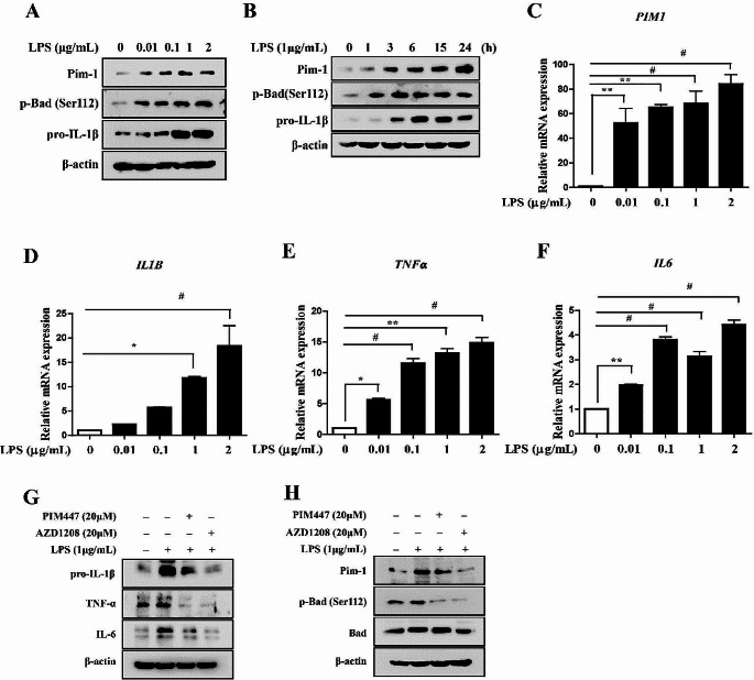
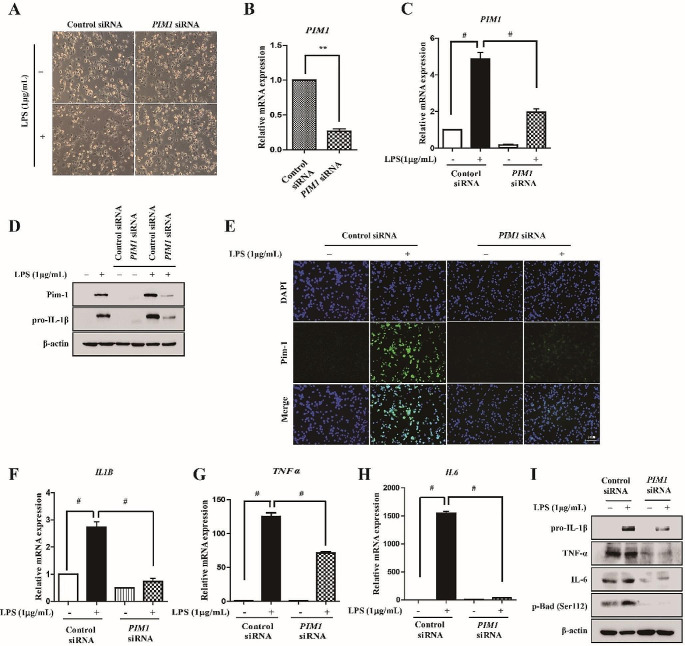
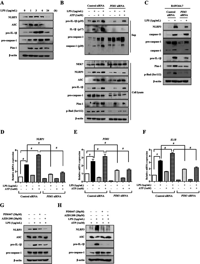
Lipopolysaccharide (LPS) was used to induce inflammation, followed by PIM1 knockdown. Western blot, immunoprecipitation, immunofluorescence, and RT-PCR assays were used to assess the effect of PIM1 knockdown on LPS-induced inflammation.
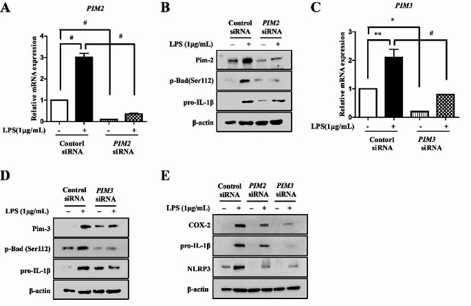
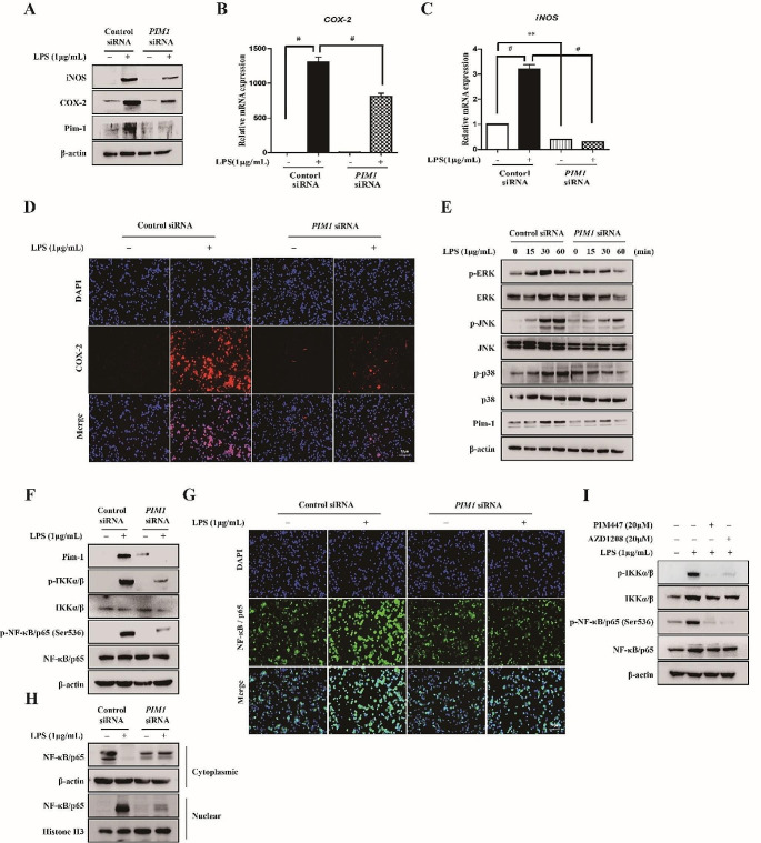
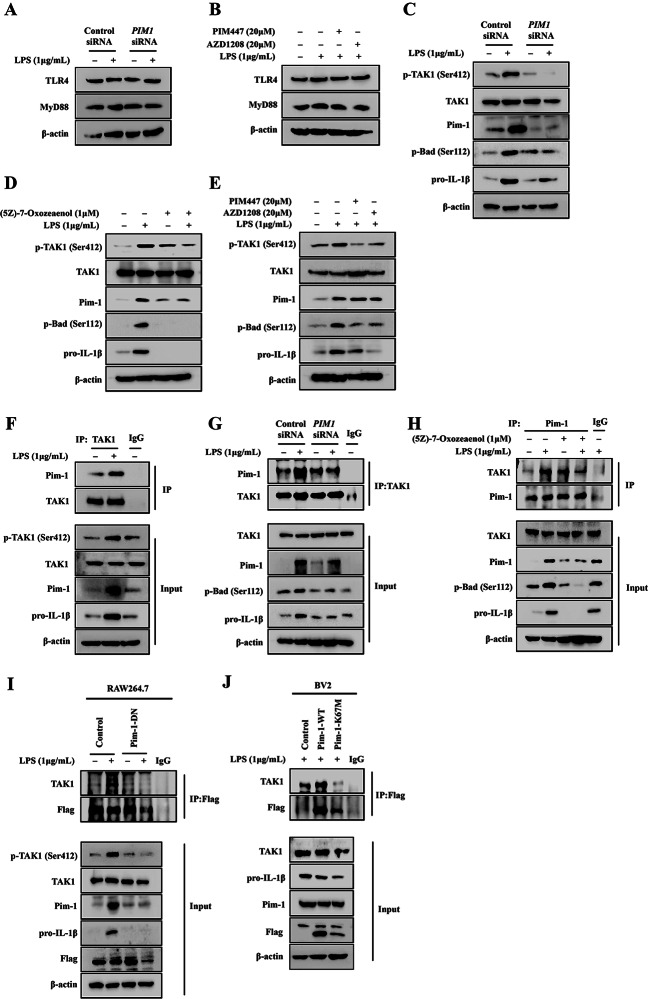
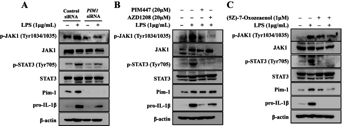
PIM1 knockdown in macrophage-like THP-1 cells suppressed LPS-induced upregulation of pro-inflammatory cytokines, inducible nitric oxide synthase, cyclooxygenase-2, phosphorylated Janus kinase, signal transducer and activator of transcription 3, extracellular signal-regulated kinase, c-Jun N-terminal kinase, p38, and nuclear factor kappa B p65 (NF-κB p65). It also suppressed upregulation of inhibitor of NF-κB kinase α/β and enhanced the nuclear translocation of NF-κB p65. Moreover, it inhibited the upregulation of Nod-like receptor family pyrin domain-containing 3 (NLRP3) and cleavage of caspase-1 induced by co-treatment of LPS with adenosine triphosphate. Additionally, p-transforming growth factor-β-activated kinase 1 (TAK1) interacted with Pim-1. All three members of Pim kinases (Pim-1, Pim-2, and Pim-3) were required for LPS-mediated inflammation in macrophages; however, unlike Pim-1 and Pim-3, Pim-2 functioned as a negative regulator of T cell activity.
Pim-1 interacts with TAK1 in LPS-induced inflammatory responses and is involved in MAPK/NF-κB/NLRP3 signaling pathways. Additionally, considering the negative regulatory role of Pim-2 in T cells, further in-depth studies on their respective functions are needed.
本观察性研究旨在探讨 Pim-1 在炎症信号通路中的调节机制。
使用了 THP-1、RAW 264.7、BV2 和 Jurkat 人 T 细胞系。
无。
使用脂多糖(LPS)诱导炎症,然后敲低 PIM1。使用 Western blot、免疫沉淀、免疫荧光和 RT-PCR 检测 PIM1 敲低对 LPS 诱导炎症的影响。
巨噬细胞样 THP-1 细胞中 PIM1 的敲低抑制了 LPS 诱导的促炎细胞因子、诱导型一氧化氮合酶、环氧化酶-2、磷酸化 Janus 激酶、信号转导和转录激活因子 3、细胞外信号调节激酶、c-Jun N 末端激酶、p38 和核因子 kappa B p65(NF-κB p65)的上调。它还抑制了 NF-κB 激酶α/β抑制剂的上调,并增强了 NF-κB p65 的核转位。此外,它抑制了 LPS 与三磷酸腺苷共同处理诱导的 Nod 样受体家族吡啶结构域包含 3(NLRP3)的上调和半胱天冬酶-1 的切割。此外,p-转化生长因子-β激活激酶 1(TAK1)与 Pim-1 相互作用。三种 Pim 激酶(Pim-1、Pim-2 和 Pim-3)都需要在巨噬细胞中进行 LPS 介导的炎症反应;然而,与 Pim-1 和 Pim-3 不同,Pim-2 作为 T 细胞活性的负调节剂发挥作用。
Pim-1 在 LPS 诱导的炎症反应中与 TAK1 相互作用,并参与 MAPK/NF-κB/NLRP3 信号通路。此外,鉴于 Pim-2 在 T 细胞中的负调节作用,需要进一步深入研究它们各自的功能。