Department of Psychiatry, School of Clinical Medicine, University of Cambridge, Cambridge, UK.
Barcelonaβeta Brain Research Center (BBRC), Pasqual Maragall Foundation, Barcelona, Spain.
Hum Brain Mapp. 2024 Aug 1;45(11):e26798. doi: 10.1002/hbm.26798.
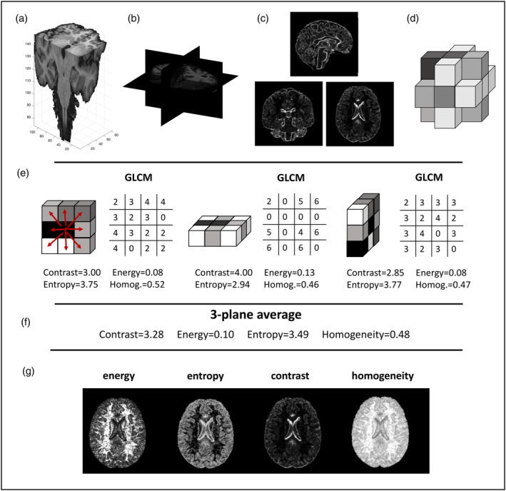
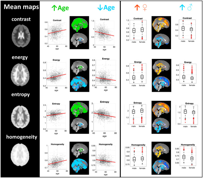
Brain atrophy and cortical thinning are typically observed in people with Alzheimer's disease (AD) and, to a lesser extent, in those with mild cognitive impairment. In asymptomatic middle-aged apolipoprotein ε4 (ΑPOE4) carriers, who are at higher risk of future AD, study reports are discordant with limited evidence of brain structural differences between carriers and non-carriers of the ε4 allele. Alternative imaging markers with higher sensitivity at the presymptomatic stage, ideally quantified using typically acquired structural MRI scans, would thus be of great benefit for the detection of early disease, disease monitoring and subject stratification. In the present cross-sectional study, we investigated textural properties of T1-weighted 3T MRI scans in relation to APOE4 genotype, age and sex. We pooled together data from the PREVENT-Dementia and ALFA studies focused on midlife healthy populations with dementia risk factors (analysable cohort: 1585 participants; mean age 56.2 ± 7.4 years). Voxel-based and texture (examined features: contrast, entropy, energy, homogeneity) based morphometry was used to identify areas of volumetric and textural differences between APOE4 carriers and non-carriers. Textural maps were generated and were subsequently harmonised using voxel-wise COMBAT. For all analyses, APOE4, sex, age and years of education were used as model predictors. Interactions between APOE4 and age were further examined. There were no group differences in regional brain volume or texture based on APOE4 carriership or when age × APOE4 interactions were examined. Older people tended to have a less homogeneous textural profile in grey and white matter and a more homogeneous profile in the ventricles. A more heterogeneous textural profile was observed for females in areas such as the ventricles, frontal and parietal lobes and for males in the brainstem, cerebellum, precuneus and cingulate. Overall, we have shown the absence of volumetric and textural differences between APOE4 carriers and non-carriers at midlife and have established associations of textural features with ageing and sex.
大脑萎缩和皮质变薄通常在阿尔茨海默病(AD)患者中观察到,在认知障碍程度较轻的患者中也会观察到。在无症状的中年载脂蛋白 E4(APOE4)携带者中,他们未来患 AD 的风险更高,但研究报告显示,携带和不携带 ε4 等位基因的携带者之间的脑结构差异存在分歧,证据有限。在无症状阶段具有更高敏感性的替代成像标志物,理想情况下使用通常获得的结构 MRI 扫描进行定量,对于早期疾病的检测、疾病监测和受试者分层将非常有益。在本横断面研究中,我们研究了 T1 加权 3T MRI 扫描的纹理特性与 APOE4 基因型、年龄和性别之间的关系。我们将重点关注有痴呆风险因素的中年健康人群的 PREVENT-Dementia 和 ALFA 研究的数据汇集在一起(可分析队列:1585 名参与者;平均年龄 56.2±7.4 岁)。体素和纹理(检查特征:对比度、熵、能量、同质性)形态计量学用于识别 APOE4 携带者和非携带者之间的体积和纹理差异区域。生成纹理图谱,并使用体素级 COMBAT 对其进行协调。在所有分析中,APOE4、性别、年龄和受教育年限被用作模型预测因子。进一步检查了 APOE4 与年龄之间的相互作用。基于 APOE4 携带状态或当检查年龄×APOE4 相互作用时,在区域脑容量或纹理方面没有组间差异。老年人的灰质和白质纹理模式往往不太均匀,脑室的纹理模式更均匀。女性在脑室、额叶和顶叶以及男性在脑干、小脑、楔前叶和扣带回等区域的纹理模式更为复杂。总的来说,我们表明在中年时,APOE4 携带者和非携带者之间没有体积和纹理差异,并建立了纹理特征与年龄和性别的关联。