Yokota Osamu, Miki Tomoko, Nakashima-Yasuda Hanae, Ishizu Hideki, Haraguchi Takashi, Ikeda Chikako, Hasegawa Masato, Miyashita Akinori, Ikeuchi Takeshi, Nishikawa Naoto, Takenoshita Shintaro, Sudo Koichiro, Terada Seishi, Takaki Manabu
Department of Neuropsychiatry, Okayama University Graduate School of Medicine, Dentistry and Pharmaceutical Sciences, 2-5-1 Shikata-cho, Okayama, 700-8558, Japan.
Okayama University Medical School, Okayama, Japan.
Acta Neuropathol Commun. 2024 Jul 31;12(1):121. doi: 10.1186/s40478-024-01828-6.
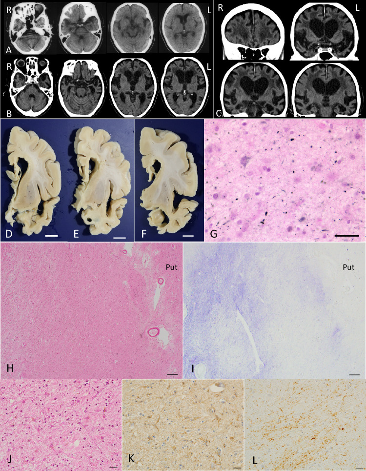
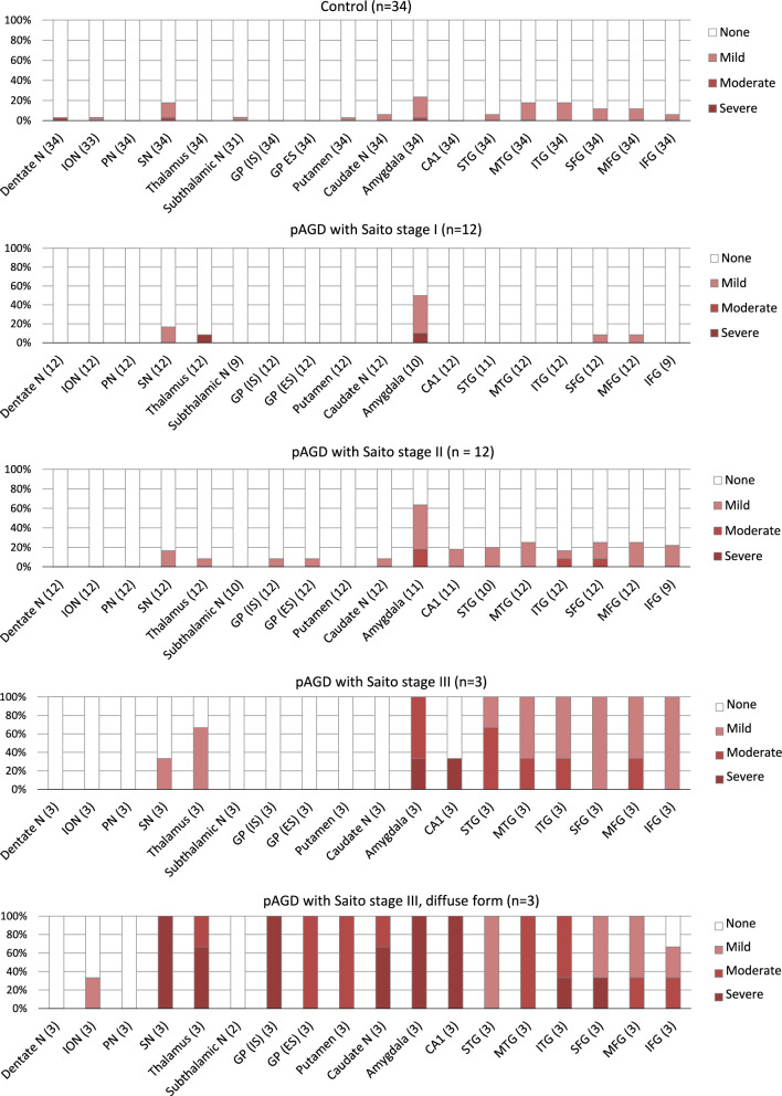
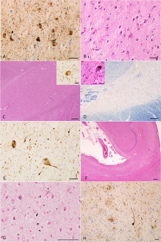
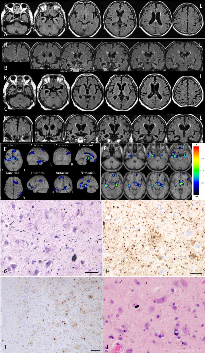
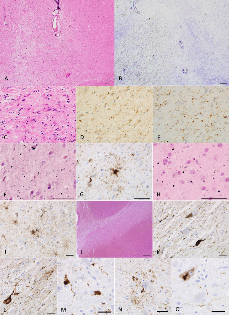
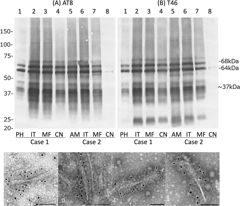
Agyrophilic grains (AGs) are age-related limbic-predominant lesions in which four-repeat tau is selectively accumulated. Because previous methodologically heterogeneous studies have demonstrated inconsistent findings on the relationship between AGs and dementia, whether AGs affect cognitive function remains unclear. To address this question, we first comprehensively evaluated the distribution and quantity of Gallyas-positive AGs and the severity of neuronal loss in the limbic, neocortical, and subcortical regions in 30 cases of pure argyrophilic grain disease (pAGD) in Braak stages I-IV and without other degenerative diseases, and 34 control cases that had only neurofibrillary tangles with Braak stages I-IV and no or minimal Aβ deposits. Then, we examined whether AGs have independent effects on neuronal loss and dementia by employing multivariate ordered logistic regression and binomial logistic regression. Of 30 pAGD cases, three were classified in diffuse form pAGD, which had evident neuronal loss not only in the limbic region but also in the neocortex and subcortical nuclei. In all 30 pAGD cases, neuronal loss developed first in the amygdala, followed by temporo-frontal cortex, hippocampal CA1, substantia nigra, and finally, the striatum and globus pallidus with the progression of Saito AG stage. In multivariate analyses of 30 pAGD and 34 control cases, the Saito AG stage affected neuronal loss in the amygdala, hippocampal CA1, temporo-frontal cortex, striatum, globus pallidus, and substantia nigra independent of the age, Braak stage, and limbic-predominant age-related TDP-43 encephalopathy (LATE-NC) stage. In multivariate analyses of 23 pAGD and 28 control cases that lacked two or more lacunae and/or one or more large infarctions, 100 or more AGs per × 400 visual field in the amygdala (OR 10.02, 95% CI 1.12-89.43) and hippocampal CA1 (OR 12.22, 95% CI 1.70-87.81), and the presence of AGs in the inferior temporal cortex (OR 8.18, 95% CI 1.03-65.13) affected dementia independent of age, moderate Braak stages (III-IV), and LATE-NC. Given these findings, the high density of limbic AGs and the increase of AGs in the inferior temporal gyrus may contribute to the occurrence of dementia through neuronal loss, at least in cases in a low to moderate Braak stage.
嗜银颗粒(AGs)是与年龄相关的以边缘系统为主的病变,其中四重复tau蛋白选择性积累。由于先前方法学上异质性的研究在AGs与痴呆症之间的关系上得出了不一致的结果,AGs是否影响认知功能仍不清楚。为了解决这个问题,我们首先全面评估了30例Braak分期I-IV期且无其他退行性疾病的纯嗜银颗粒病(pAGD)病例以及34例仅伴有Braak分期I-IV期神经原纤维缠结且无或仅有少量Aβ沉积的对照病例中,Gallyas阳性AGs在边缘系统、新皮质和皮质下区域的分布和数量,以及神经元丢失的严重程度。然后,我们通过多变量有序逻辑回归和二项逻辑回归分析,研究AGs是否对神经元丢失和痴呆有独立影响。在30例pAGD病例中,3例被归类为弥漫型pAGD,其不仅在边缘系统区域,而且在新皮质和皮质下核团都有明显的神经元丢失。在所有30例pAGD病例中,随着Saito AG分期的进展,神经元丢失首先发生在杏仁核,随后是颞额叶皮质、海马CA1区、黑质,最后是纹状体和苍白球。在对30例pAGD病例和34例对照病例的多变量分析中,Saito AG分期独立于年龄、Braak分期和边缘系统为主的年龄相关TDP-43脑病(LATE-NC)分期,影响杏仁核、海马CA1区、颞额叶皮质、纹状体、苍白球和黑质中的神经元丢失。在对23例pAGD病例和28例对照病例(这些病例没有两个或更多腔隙和/或一个或更多大梗死灶)的多变量分析中,杏仁核(比值比10.02,95%可信区间1.12-89.43)和海马CA1区(比值比12.22,95%可信区间1.70-87.81)每×400视野中100个或更多AGs,以及颞下回中AGs的存在(比值比8.18,95%可信区间1.03-65.13)独立于年龄、中度Braak分期(III-IV期)和LATE-NC影响痴呆。基于这些发现,边缘系统AGs的高密度以及颞下回中AGs的增加可能至少在低至中度Braak分期的病例中通过神经元丢失导致痴呆的发生。