• 文献检索
  • 文档翻译
  • 深度研究
  • 学术资讯
  • Suppr Zotero 插件Zotero 插件
  • 邀请有礼
  • 套餐&价格
  • 历史记录
应用&插件
Suppr Zotero 插件Zotero 插件浏览器插件Mac 客户端Windows 客户端微信小程序
定价
高级版会员购买积分包购买API积分包
服务
文献检索文档翻译深度研究API 文档MCP 服务
关于我们
关于 Suppr公司介绍联系我们用户协议隐私条款
关注我们

Suppr 超能文献

核心技术专利:CN118964589B侵权必究
粤ICP备2023148730 号-1Suppr @ 2026

文献检索

告别复杂PubMed语法,用中文像聊天一样搜索,搜遍4000万医学文献。AI智能推荐,让科研检索更轻松。

立即免费搜索

文件翻译

保留排版,准确专业,支持PDF/Word/PPT等文件格式,支持 12+语言互译。

免费翻译文档

深度研究

AI帮你快速写综述,25分钟生成高质量综述,智能提取关键信息,辅助科研写作。

立即免费体验

发育中眼睛的时间转录组分析

Temporal Transcriptomic Profiling of the Developing Eye.

作者信息

Hack Samantha J, Petereit Juli, Tseng Kelly Ai-Sun

机构信息

Department of Biological Sciences, Western Michigan University, Kalamazoo, MI 49008, USA.

Nevada Bioinformatics Center, University of Nevada, Reno.

出版信息

bioRxiv. 2024 Jul 22:2024.07.20.603187. doi: 10.1101/2024.07.20.603187.

DOI:10.1101/2024.07.20.603187
PMID:39091861
原文链接:https://pmc.ncbi.nlm.nih.gov/articles/PMC11291033/
Abstract

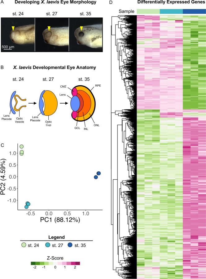
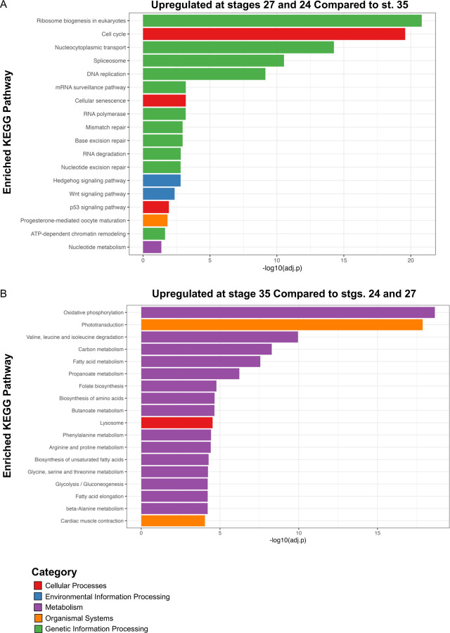
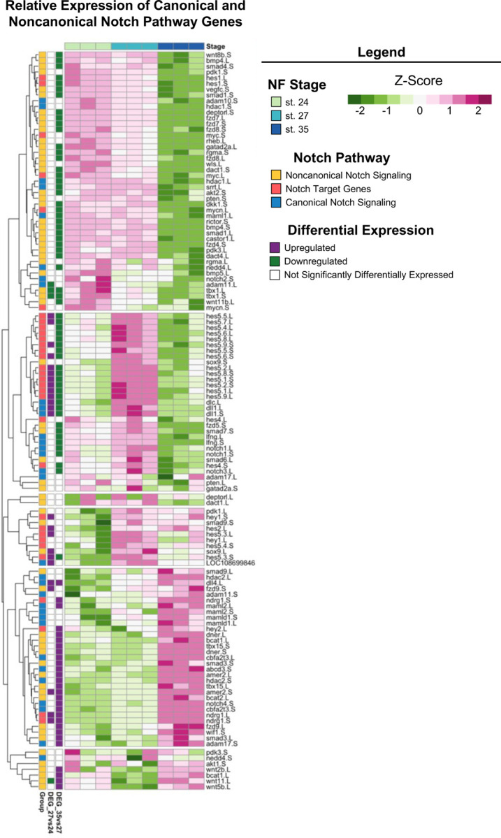
Retinal progenitor cells (RPCs) are a multipotent and highly proliferative population that give rise to all retinal cell types during organogenesis. Defining their molecular signature is a key step towards identifying suitable approaches to treat visual impairments. Here, we performed RNA-sequencing of whole eyes from at three embryonic stages and used differential expression analysis to define the transcriptomic profiles of optic tissues containing proliferating and differentiating RPCs during retinogenesis. Gene Ontology and KEGG pathway analyses showed that genes associated with developmental pathways (including Wnt and Hedgehog signaling) were upregulated during the period of active RPC proliferation in early retinal development (Nieuwkoop Faber st. 24 and 27). Developing eyes had dynamic expression profiles and shifted to enrichment for metabolic processes and phototransduction during RPC progeny specification and differentiation (st. 35). Furthermore, conserved adult eye regeneration genes were also expressed during early retinal development including , , , and Notch signaling components. The eye transcriptomic profiles presented here span RPC proliferation to retinogenesis and included regrowth-competent stages. Thus, our dataset provides a rich resource to uncover molecular regulators of RPC activity and will allow future studies to address regulators of RPC proliferation during eye repair and regrowth.

摘要

视网膜祖细胞(RPCs)是一类多能且高度增殖的细胞群体,在器官发生过程中可产生所有视网膜细胞类型。确定它们的分子特征是找到治疗视力障碍合适方法的关键一步。在此,我们对三个胚胎阶段的全眼进行了RNA测序,并使用差异表达分析来确定视网膜发育过程中包含增殖和分化RPCs的视神经组织的转录组图谱。基因本体论和KEGG通路分析表明,在视网膜早期发育(Nieuwkoop Faber第24和27阶段)RPC活跃增殖期间,与发育途径(包括Wnt和Hedgehog信号通路)相关的基因上调。在RPC子代特化和分化过程(第35阶段),发育中的眼睛具有动态表达谱,并转向富集代谢过程和光转导相关基因。此外,包括、、和Notch信号成分在内的保守的成年眼再生基因在视网膜早期发育过程中也有表达。这里呈现的眼睛转录组图谱涵盖了RPC增殖到视网膜形成的过程,包括具备再生能力的阶段。因此,我们的数据集为揭示RPC活性的分子调节因子提供了丰富资源,并将使未来的研究能够探讨眼睛修复和再生过程中RPC增殖的调节因子。

https://cdn.ncbi.nlm.nih.gov/pmc/blobs/6ec1/11291033/bf255241ecf3/nihpp-2024.07.20.603187v1-f0007.jpg
https://cdn.ncbi.nlm.nih.gov/pmc/blobs/6ec1/11291033/368c057b7f8f/nihpp-2024.07.20.603187v1-f0001.jpg
https://cdn.ncbi.nlm.nih.gov/pmc/blobs/6ec1/11291033/9e0c653884cb/nihpp-2024.07.20.603187v1-f0002.jpg
https://cdn.ncbi.nlm.nih.gov/pmc/blobs/6ec1/11291033/03812e201e0e/nihpp-2024.07.20.603187v1-f0003.jpg
https://cdn.ncbi.nlm.nih.gov/pmc/blobs/6ec1/11291033/6917a4a96a7d/nihpp-2024.07.20.603187v1-f0004.jpg
https://cdn.ncbi.nlm.nih.gov/pmc/blobs/6ec1/11291033/c8af9597c840/nihpp-2024.07.20.603187v1-f0005.jpg
https://cdn.ncbi.nlm.nih.gov/pmc/blobs/6ec1/11291033/cb25463d36d7/nihpp-2024.07.20.603187v1-f0006.jpg
https://cdn.ncbi.nlm.nih.gov/pmc/blobs/6ec1/11291033/bf255241ecf3/nihpp-2024.07.20.603187v1-f0007.jpg

相似文献

1
Temporal Transcriptomic Profiling of the Developing Eye.发育中眼睛的时间转录组分析
bioRxiv. 2024 Jul 22:2024.07.20.603187. doi: 10.1101/2024.07.20.603187.
2
Temporal Transcriptomic Profiling of the Developing Eye.发育中眼睛的时间转录组分析。
Cells. 2024 Aug 21;13(16):1390. doi: 10.3390/cells13161390.
3
Studying In Vivo Retinal Progenitor Cell Proliferation in Xenopus laevis.研究非洲爪蟾体内视网膜祖细胞的增殖
Methods Mol Biol. 2020;2092:19-33. doi: 10.1007/978-1-0716-0175-4_2.
4
Single-Cell RNA Sequencing of hESC-Derived 3D Retinal Organoids Reveals Novel Genes Regulating RPC Commitment in Early Human Retinogenesis.人胚胎干细胞来源的 3D 视网膜类器官的单细胞 RNA 测序揭示了早期人视网膜发生中调控 RPC 定型的新基因。
Stem Cell Reports. 2019 Oct 8;13(4):747-760. doi: 10.1016/j.stemcr.2019.08.012. Epub 2019 Sep 19.
5
Gene expression is dynamically regulated in retinal progenitor cells prior to and during overt cellular differentiation.在明显的细胞分化之前及分化过程中,视网膜祖细胞中的基因表达受到动态调控。
Gene Expr Patterns. 2014 Jan;14(1):42-54. doi: 10.1016/j.gep.2013.10.003. Epub 2013 Oct 19.
6
Notch Is Required for Neural Progenitor Proliferation During Embryonic Eye Regrowth.Notch在胚胎眼睛再生过程中对神经祖细胞增殖是必需的。
Int J Mol Sci. 2025 Mar 14;26(6):2637. doi: 10.3390/ijms26062637.
7
V-ATPase Regulates Retinal Progenitor Cell Proliferation During Eye Regrowth in .V-ATPase 在 . 中调控眼再生过程中的视网膜祖细胞增殖
J Ocul Pharmacol Ther. 2023 Oct;39(8):499-508. doi: 10.1089/jop.2022.0085. Epub 2023 Mar 3.
8
Laminin β2 Chain Regulates Retinal Progenitor Cell Mitotic Spindle Orientation via Dystroglycan.层粘连蛋白β2 链通过 dystroglycan 调节视网膜祖细胞有丝分裂纺锤体取向。
J Neurosci. 2018 Jun 27;38(26):5996-6010. doi: 10.1523/JNEUROSCI.0551-18.2018. Epub 2018 May 31.
9
Temporal Requirement of Mab21l2 During Eye Development in Chick Reveals Stage-Dependent Functions for Retinogenesis.Mab21l2 在鸡眼发育过程中的时间需求揭示了其对视网膜发生的阶段依赖性功能。
Invest Ophthalmol Vis Sci. 2018 Aug 1;59(10):3869-3878. doi: 10.1167/iovs.18-24236.
10
Transcriptome of Atoh7 retinal progenitor cells identifies new Atoh7-dependent regulatory genes for retinal ganglion cell formation.Atoh7视网膜祖细胞的转录组鉴定出视网膜神经节细胞形成中依赖Atoh7的新调控基因。
Dev Neurobiol. 2014 Nov;74(11):1123-40. doi: 10.1002/dneu.22188. Epub 2014 May 22.

本文引用的文献

1
Notch Is Required for Neural Progenitor Proliferation During Embryonic Eye Regrowth.Notch在胚胎眼睛再生过程中对神经祖细胞增殖是必需的。
Int J Mol Sci. 2025 Mar 14;26(6):2637. doi: 10.3390/ijms26062637.
2
Kdm7a expression is spatiotemporally regulated in developing Xenopus laevis embryos, and its overexpression influences late retinal development.Kdm7a的表达在非洲爪蟾胚胎发育过程中受到时空调控,其过表达会影响视网膜后期发育。
Dev Dyn. 2024 May;253(5):508-518. doi: 10.1002/dvdy.670. Epub 2023 Nov 1.
3
Cell-type expression and activation by light of neuropsins in the developing and mature retina.
视紫红质在发育中和成熟视网膜中的细胞类型表达及光激活情况。
Front Cell Neurosci. 2023 Sep 20;17:1266945. doi: 10.3389/fncel.2023.1266945. eCollection 2023.
4
The Gene Ontology knowledgebase in 2023.2023 版基因本体论知识库。
Genetics. 2023 May 4;224(1). doi: 10.1093/genetics/iyad031.
5
Notch signaling in development and homeostasis.Notch 信号通路在发育和稳态中的作用。
Development. 2023 Feb 15;150(4). doi: 10.1242/dev.201138. Epub 2023 Feb 16.
6
Xenbase: key features and resources of the Xenopus model organism knowledgebase.Xenbase:爪蟾模式生物知识库的主要特点和资源
Genetics. 2023 May 4;224(1). doi: 10.1093/genetics/iyad018.
7
Publisher Correction: Cell landscape of larval and adult Xenopus laevis at single-cell resolution.出版商更正:非洲爪蟾幼体和成体在单细胞分辨率下的细胞图谱。
Nat Commun. 2022 Sep 1;13(1):5142. doi: 10.1038/s41467-022-32733-y.
8
Cell fate decisions, transcription factors and signaling during early retinal development.早期视网膜发育过程中的细胞命运决定、转录因子和信号转导。
Prog Retin Eye Res. 2022 Nov;91:101093. doi: 10.1016/j.preteyeres.2022.101093. Epub 2022 Jul 8.
9
The Xenopus phenotype ontology: bridging model organism phenotype data to human health and development.非洲爪蟾表型本体:连接模型生物表型数据与人类健康和发育。
BMC Bioinformatics. 2022 Mar 22;23(1):99. doi: 10.1186/s12859-022-04636-8.
10
Notch signaling enhances bone regeneration in the zebrafish mandible.Notch 信号通路增强斑马鱼下颌骨的再生。
Development. 2022 Mar 1;149(5). doi: 10.1242/dev.199995. Epub 2022 Mar 11.