Banu Jahira, Dhakshnamoorthy Nithya, Sakthivel Sulochana
Anatomy, Jawaharlal Institute of Postgraduate Medical Education and Research, Pondicherry, IND.
Cureus. 2024 Jul 10;16(7):e64282. doi: 10.7759/cureus.64282. eCollection 2024 Jul.
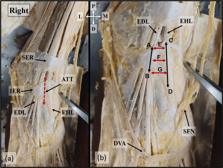
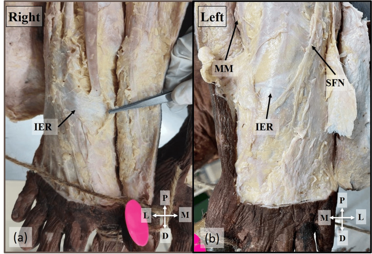
Anterior tarsal tunnel (ATT) syndrome is caused by the compression of the deep fibular nerve (DFN) within the ATT beneath the inferior extensor retinaculum, bounded by the tendons of the extensor hallucis longus (EHL) and extensor digitorum longus (EDL). Compression may result from direct trauma, repetitive mechanical irritation, and thrombosis of the dorsalis pedis artery. Injury to the contents of ATT could occur during ankle arthroscopy. Therefore, this study was undertaken to provide a detailed description of the anatomy of the ATT and its clinical implications.
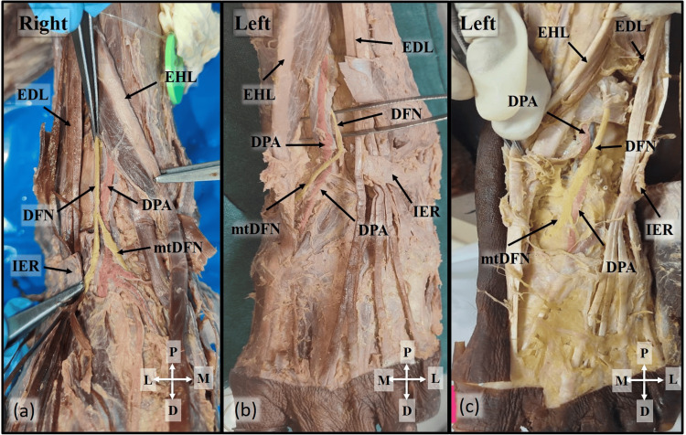
Ten formalin-fixed cadavers were utilized for the study. The ATT was identified between the tendons of the EHL and EDL. The length at the medial and lateral boundaries and the width at the proximal end, middle, and distal end of the ATT were measured using a digital Vernier calliper.
The mean length of the medial border of the tunnel was 31.42±8.44 mm, while the lateral border was 20.39±4.39 mm. The width of the ATT increased from the proximal to the distal end. DFN was related to the DPA laterally in 15 limbs and medially in five limbs within the tunnel.
The present study not only describes the intricate anatomy of the ATT but also describes the patterns of DFN and DPA within the tunnel. Understanding the anatomy of ATT is crucial, as it paves the way for safe and efficient surgical interventions, thereby significantly reducing the risk of neurovascular damage during surgical procedures.
跗骨前隧道(ATT)综合征是由位于下伸肌支持带下方的ATT内的腓深神经(DFN)受压引起的,该区域由拇长伸肌(EHL)和趾长伸肌(EDL)的肌腱界定。压迫可能由直接创伤、重复性机械刺激和足背动脉血栓形成引起。踝关节镜检查期间可能会损伤ATT内的结构。因此,本研究旨在详细描述ATT的解剖结构及其临床意义。
本研究使用了10具福尔马林固定的尸体。在EHL和EDL的肌腱之间识别出ATT。使用数字游标卡尺测量ATT内侧和外侧边界的长度以及近端、中间和远端的宽度。
隧道内侧边界的平均长度为31.42±8.44毫米,而外侧边界为20.39±4.39毫米。ATT的宽度从近端到远端逐渐增加。在隧道内,DFN在15条肢体中与足背动脉(DPA)外侧相关,在5条肢体中与DPA内侧相关。
本研究不仅描述了ATT复杂的解剖结构,还描述了隧道内DFN和DPA的分布模式。了解ATT的解剖结构至关重要,因为它为安全有效的手术干预铺平了道路,从而显著降低手术过程中神经血管损伤的风险。