Department of Neurosurgery, Graduate School of Biomedical and Health Sciences, Hiroshima University, 1-2-3 Kasumi, Minami-ku, Hiroshima-city, Hiroshima, 734-8551, Japan.
Department of Clinical Radiology, Hiroshima University Hospital, Hiroshima, Japan.
J Neurooncol. 2024 Nov;170(2):429-436. doi: 10.1007/s11060-024-04794-0. Epub 2024 Aug 12.
The T2-FLAIR mismatch sign is a characteristic imaging biomarker for astrocytoma, isocitrate dehydrogenase (IDH)-mutant. However, investigators have provided varying interpretations of the positivity/negativity of this sign given for individual cases the nature of qualitative visual assessment. Moreover, MR sequence parameters also influence the appearance of the T2-FLAIR mismatch sign. To resolve these issues, we used synthetic MR technique to quantitatively evaluate and differentiate astrocytoma from oligodendroglioma.
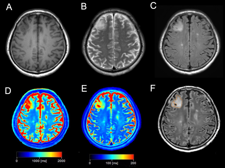
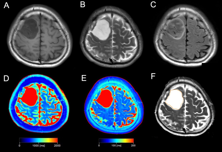
This study included 20 patients with newly diagnosed non-enhanced IDH-mutant diffuse glioma who underwent preoperative synthetic MRI using the Quantification of Relaxation Times and Proton Density by Multiecho acquisition of a saturation-recovery using Turbo spin-Echo Readout (QRAPMASTER) sequence at our institution. Two independent reviewers evaluated preoperative conventional MR images to determine the presence or absence of the T2-FLAIR mismatch sign. Synthetic MRI was used to measure T1, T2 and proton density (PD) values in the tumor lesion. Receiver operating characteristic (ROC) curve analysis was performed to evaluate the diagnostic performance.
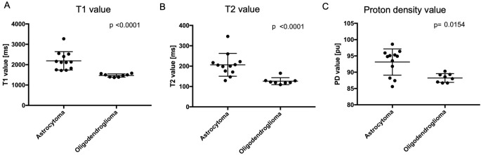
The pathological diagnoses included astrocytoma, IDH-mutant (n = 12) and oligodendroglioma, IDH-mutant and 1p/19q-codeleted (n = 8). The sensitivity and specificity of T2-FLAIR mismatch sign for astrocytoma were 66.7% and 100% [area under the ROC curve (AUC) = 0.833], respectively. Astrocytoma had significantly higher T1, T2, and PD values than did oligodendroglioma (p < 0.0001, < 0.0001, and 0.0154, respectively). A cutoff lesion T1 value of 1580 ms completely differentiated astrocytoma from oligodendroglioma (AUC = 1.00).
Quantitative evaluation of non-enhanced IDH-mutant diffuse glioma using synthetic MRI allowed for better differentiation between astrocytoma and oligodendroglioma than did conventional T2-FLAIR mismatch sign. Measurement of T1 and T2 value by synthetic MRI could improve the differentiation of IDH-mutant diffuse gliomas.
T2-FLAIR 不匹配征象是 IDH 突变型星形细胞瘤的一种特征性影像学生物标志物。然而,由于定性视觉评估的性质,研究人员对该征象的阳性/阴性给出了不同的解释。此外,MR 序列参数也会影响 T2-FLAIR 不匹配征象的表现。为了解决这些问题,我们使用合成 MR 技术对星形细胞瘤和少突胶质细胞瘤进行定量评估和区分。
本研究纳入了 20 例在我院行术前合成 MRI 检查的新诊断的非增强 IDH 突变型弥漫性胶质瘤患者,使用定量弛豫时间和质子密度通过多回波采集的饱和恢复 Turbo 自旋回波读出(QRAPMASTER)序列进行。两位独立的评估者评估术前常规 MR 图像以确定 T2-FLAIR 不匹配征象的存在或不存在。使用合成 MRI 测量肿瘤病变中的 T1、T2 和质子密度(PD)值。进行受试者工作特征(ROC)曲线分析以评估诊断性能。
病理诊断包括星形细胞瘤,IDH 突变型(n=12)和少突胶质细胞瘤,IDH 突变型和 1p/19q 联合缺失型(n=8)。T2-FLAIR 不匹配征象对星形细胞瘤的敏感性和特异性分别为 66.7%和 100%[ROC 曲线下面积(AUC)=0.833]。星形细胞瘤的 T1、T2 和 PD 值明显高于少突胶质细胞瘤(p<0.0001、<0.0001 和 0.0154)。病变 T1 值为 1580 ms 的截断值可完全区分星形细胞瘤和少突胶质细胞瘤(AUC=1.00)。
使用合成 MRI 对非增强 IDH 突变型弥漫性胶质瘤进行定量评估可更好地区分星形细胞瘤和少突胶质细胞瘤,优于常规 T2-FLAIR 不匹配征象。合成 MRI 测量 T1 和 T2 值可以提高 IDH 突变型弥漫性胶质瘤的鉴别能力。