Tipirneni-Sajja Aaryani, Shrestha Utsav, Esparza Juan, Morin Cara E, Kannengiesser Stephan, Roberts Nathan T, Peeters Johannes M, Sharma Samir D, Hu Houchun H
Department of Biomedical Engineering, The University of Memphis, Memphis, Tennessee, USA.
Diagnostic Imaging, St. Jude Children's Research Hospital, Memphis, Tennessee, USA.
J Magn Reson Imaging. 2025 Mar;61(3):1110-1132. doi: 10.1002/jmri.29526. Epub 2024 Aug 12.
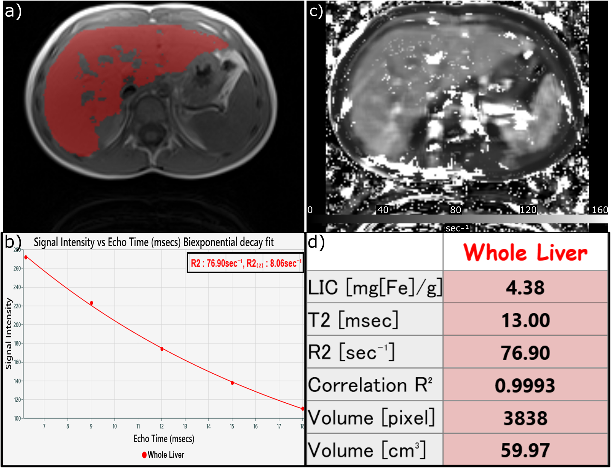
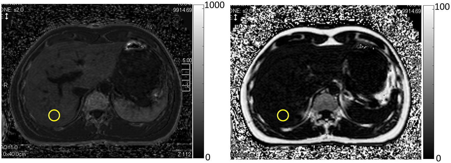
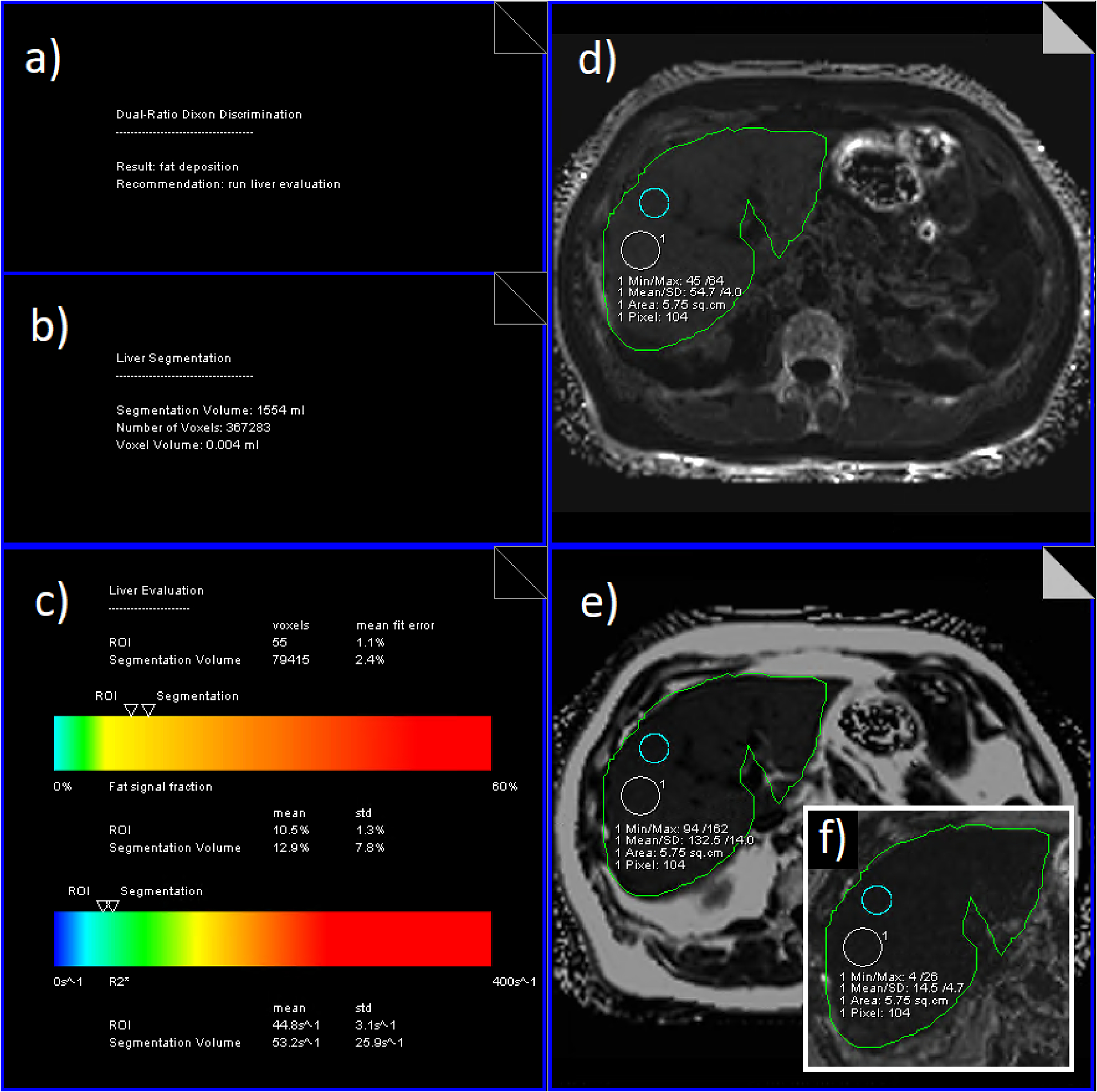
The role of MRI to estimate liver iron concentration (LIC) for identifying patients with iron overload and guiding the titration of chelation therapy is increasingly established for routine clinical practice. However, the existence of multiple MRI-based LIC quantification techniques limits standardization and widespread clinical adoption. In this article, we review the existing and widely accepted MRI-based LIC estimation methods at 1.5 T and 3 T: signal intensity ratio (SIR) and relaxometry (R2 and R2*) and discuss the basic principles, acquisition and analysis protocols, and MRI-LIC calibrations for each technique. Further, we provide an up-to-date information on MRI vendor implementations and available offline commercial and free software for each MRI-based LIC quantification approach. We also briefly review the emerging and advanced MRI techniques for LIC estimation and their current limitations for clinical use. Lastly, we discuss the implications of MRI-based LIC measurements on clinical use and decision-making in the management of patients with iron overload. Some of the key highlights from this review are as follows: 1) Both R2 and R2* can estimate accurate and reproducible LIC, when validated acquisition parameters and analysis protocols are applied, 2) Although the Ferriscan R2 method has been widely used, recent consensus and guidelines endorse R2*-MRI as the most accurate and reproducible method for LIC estimation, 3) Ongoing efforts aim to establish R2*-MRI as the standard approach for quantifying LIC, and 4) Emerging R2*-MRI techniques employ radial sampling strategies and offer improved motion compensation and broader dynamic range for LIC estimation. EVIDENCE LEVEL: 1 TECHNICAL EFFICACY: Stage 2.
磁共振成像(MRI)在评估肝脏铁浓度(LIC)以识别铁过载患者并指导螯合治疗滴定方面的作用,在常规临床实践中越来越得到认可。然而,多种基于MRI的LIC定量技术的存在限制了标准化和广泛的临床应用。在本文中,我们回顾了在1.5 T和3 T磁场强度下现有的且被广泛接受的基于MRI的LIC估计方法:信号强度比(SIR)和弛豫测量法(R2和R2*),并讨论了每种技术的基本原理、采集和分析方案以及MRI-LIC校准。此外,我们提供了关于MRI供应商实施情况以及每种基于MRI的LIC定量方法的可用离线商业软件和免费软件的最新信息。我们还简要回顾了用于LIC估计的新兴和先进MRI技术及其目前在临床应用中的局限性。最后,我们讨论了基于MRI的LIC测量对铁过载患者管理中临床应用和决策的影响。本综述的一些关键要点如下:1)当应用经过验证的采集参数和分析方案时,R2和R2都可以准确且可重复地估计LIC;2)尽管Ferriscan R2方法已被广泛使用,但最近的共识和指南认可R2 - MRI作为LIC估计最准确且可重复的方法;3)正在进行的努力旨在将R2* - MRI确立为量化LIC的标准方法;4)新兴的R2* - MRI技术采用径向采样策略,为LIC估计提供了更好的运动补偿和更宽的动态范围。证据水平:1 技术效能:2级