State Key Laboratory of Oral & Maxillofacial Reconstruction and Regeneration, Key Laboratory of Oral Biomedicine Ministry of Education, Hubei Key Laboratory of Stomatology, School & Hospital of Stomatology, Wuhan University, Luoyu Road 237, Wuhan, 430079, PR China.
Oral Histopathology Department, School and Hospital of Stomatology, Wuhan University, Wuhan, 430079, China.
Diagn Pathol. 2024 Aug 13;19(1):109. doi: 10.1186/s13000-024-01530-0.
Clear cell odontogenic carcinoma (CCOC) is an odontogenic carcinoma characterized by sheets and islands of vacuolated and clear cells. The diagnosis of atypical CCOC can pose a challenge when tumor cells deviate from their characteristic clear morphology, even with the aid of genetic profiling for CCOC identification.
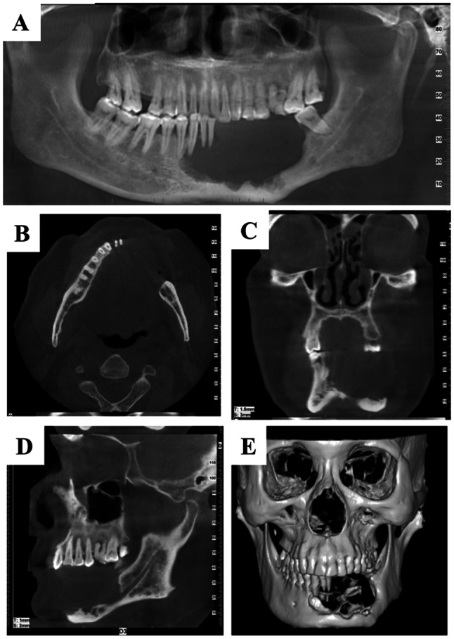
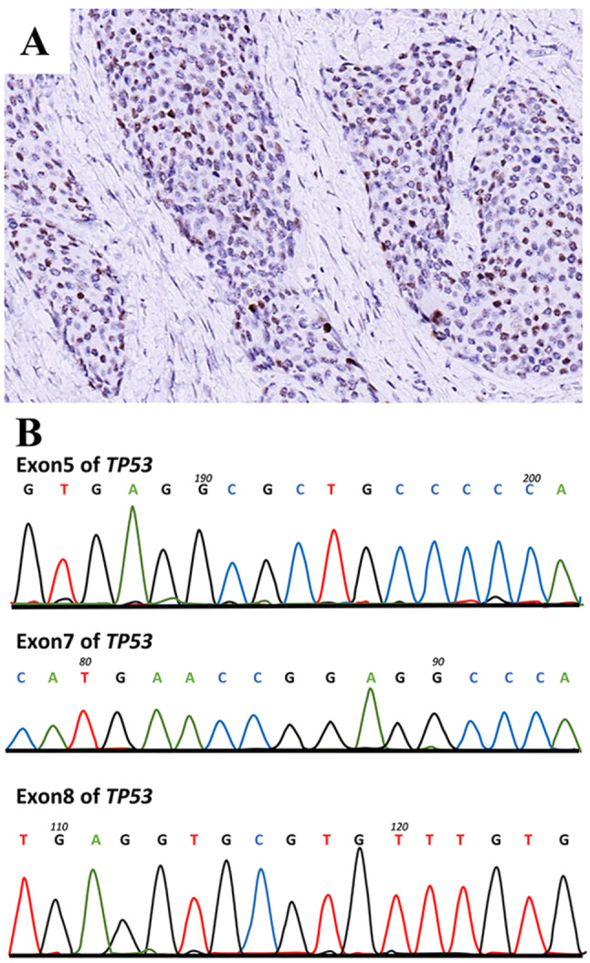
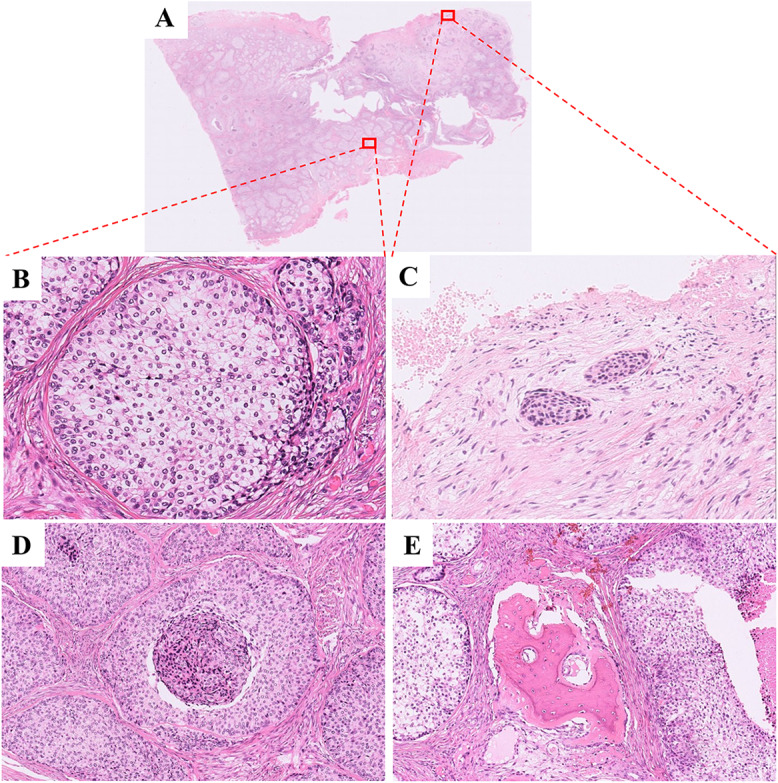
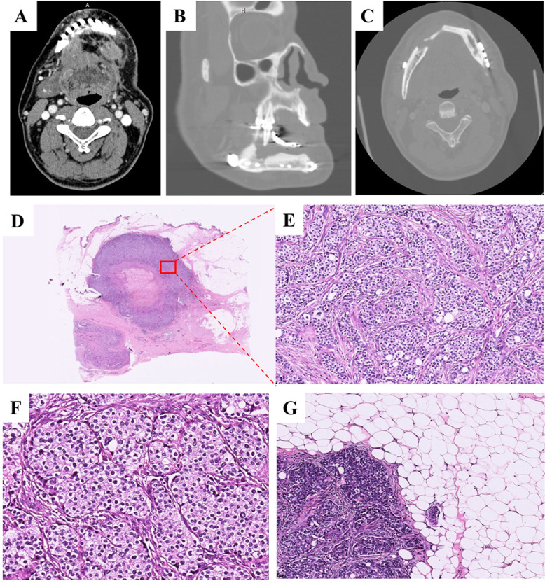
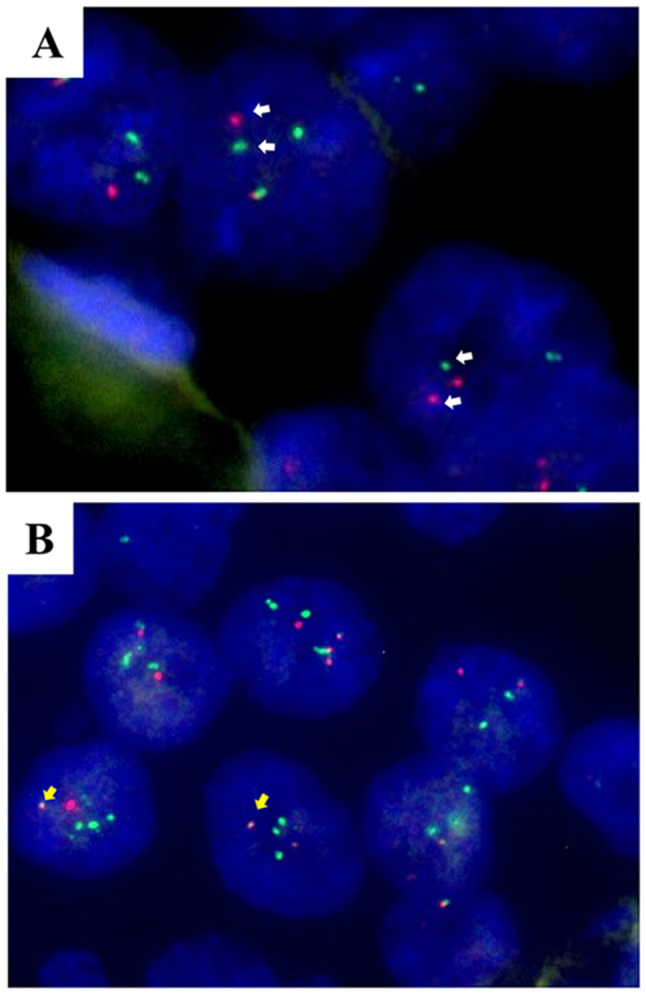
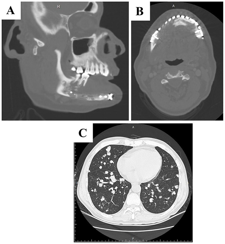
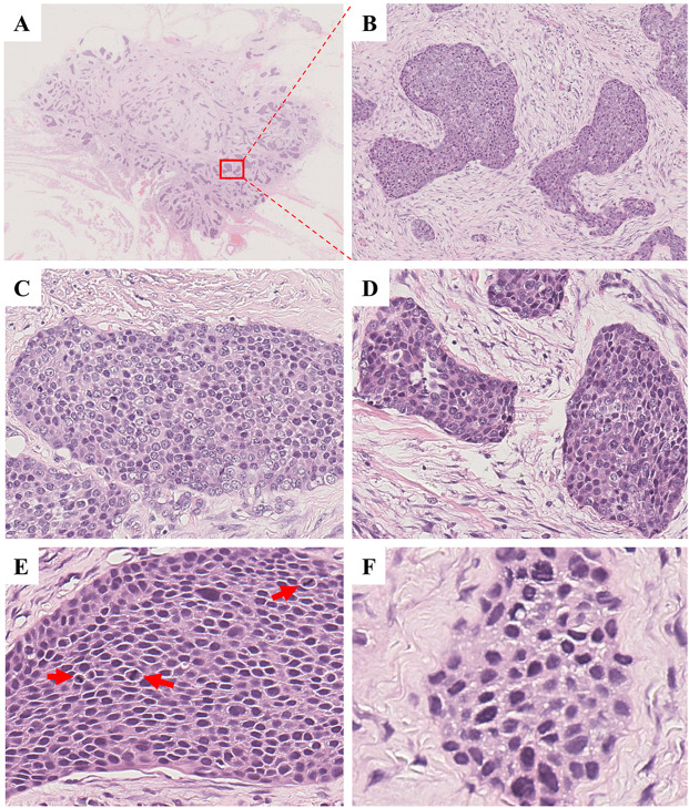
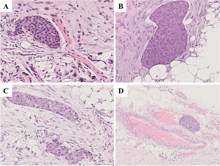
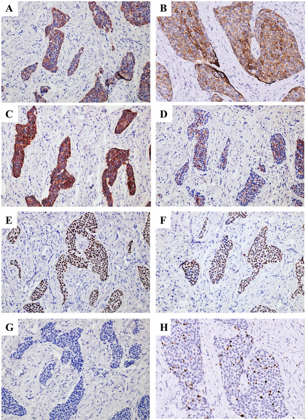
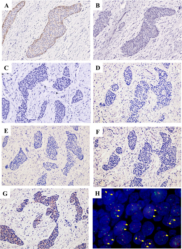
In this manuscript, we detailed the inaugural instance of a recurrently recurring clear cell odontogenic carcinoma (CCOC) with pronounced squamous differentiation in a 64-year-old male. The primary tumor in this individual initially displayed a biphasic clear cell phenotype. However, subsequent to the third recurrence, the clear tumor cells were entirely supplanted by epidermoid cells characterized by eosinophilic cytoplasm, vesicular chromatin, and prominent nucleoli. Notable aggressive attributes such as necrosis, conspicuous cytological malignancy, perineural dissemination, and vascular invasion were noted. Additionally, the tumor progressed to manifest lung metastases. The tumor cells exhibited positive immunoreactivity for AE1/AE3, KRT19, Pan-CK, EMA, P40, P63, CK34βE12, and P53, while they tested negative for CK35βH11, KRT7, S-100, and neuroendocrine markers. The Ki-67 proliferation index was calculated at an average of 15%. Furthermore, FISH analysis unveiled the presence of the EWSR1::ATF1 gene fusion.
This case illustrated a rare and aggressive case of CCOC characterized by significant squamous differentiation upon recurrence of the tumor.
透明细胞牙源性癌(CCOC)是一种牙源性癌,其特征为有空泡和透明细胞的片状和岛屿状。当肿瘤细胞偏离其特征性透明形态时,即使借助 CCOC 鉴定的基因谱分析,也可能难以诊断非典型 CCOC。
本文详细介绍了首例复发性透明细胞牙源性癌(CCOC)的病例,该病例具有明显的鳞状分化,发生于一名 64 岁男性。该患者的原发性肿瘤最初表现为双相透明细胞表型。然而,在第三次复发后,透明肿瘤细胞完全被具有嗜酸性细胞质、泡状染色质和明显核仁的表皮样细胞取代。明显的侵袭性特征,如坏死、明显的细胞学恶性、神经周围扩散和血管浸润均有记录。此外,肿瘤进展至肺转移。肿瘤细胞对 AE1/AE3、KRT19、Pan-CK、EMA、P40、P63、CK34βE12 和 P53 表现出阳性免疫反应,而对 CK35βH11、KRT7、S-100 和神经内分泌标志物呈阴性。Ki-67 增殖指数平均为 15%。此外,FISH 分析显示存在 EWSR1::ATF1 基因融合。
该病例展示了一例罕见且侵袭性的 CCOC 病例,其特征为肿瘤复发时具有显著的鳞状分化。