• 文献检索
  • 文档翻译
  • 深度研究
  • 学术资讯
  • Suppr Zotero 插件Zotero 插件
  • 邀请有礼
  • 套餐&价格
  • 历史记录
应用&插件
Suppr Zotero 插件Zotero 插件浏览器插件Mac 客户端Windows 客户端微信小程序
定价
高级版会员购买积分包购买API积分包
服务
文献检索文档翻译深度研究API 文档MCP 服务
关于我们
关于 Suppr公司介绍联系我们用户协议隐私条款
关注我们

Suppr 超能文献

核心技术专利:CN118964589B侵权必究
粤ICP备2023148730 号-1Suppr @ 2026

文献检索

告别复杂PubMed语法,用中文像聊天一样搜索,搜遍4000万医学文献。AI智能推荐,让科研检索更轻松。

立即免费搜索

文件翻译

保留排版,准确专业,支持PDF/Word/PPT等文件格式,支持 12+语言互译。

免费翻译文档

深度研究

AI帮你快速写综述,25分钟生成高质量综述,智能提取关键信息,辅助科研写作。

立即免费体验

复合树脂修复治疗牙根外吸收中卓越的骨重塑:一例报告

Exceptional bone remodeling over composite restoration in the management of external root resorption: A case report.

作者信息

Zaccheo Fabrizio, Petroni Giulia, Fiorillo Luca, Heboyan Artak, Carnevale Gianfranco, Calapaj Massimo, Cervino Gabriele, Tallarico Marco

机构信息

Department of Oral and Maxillofacial Sciences, Sapienza University of Rome, Rome, Italy.

Department of Dental Research Cell, Dr. D. Y. Patil Dental College and Hospital, Dr. D. Y. Patil Vidyapeeth, Pune, India.

出版信息

SAGE Open Med Case Rep. 2024 Aug 12;12:2050313X241269588. doi: 10.1177/2050313X241269588. eCollection 2024.

DOI:10.1177/2050313X241269588
PMID:39140030
原文链接:https://pmc.ncbi.nlm.nih.gov/articles/PMC11320676/
Abstract

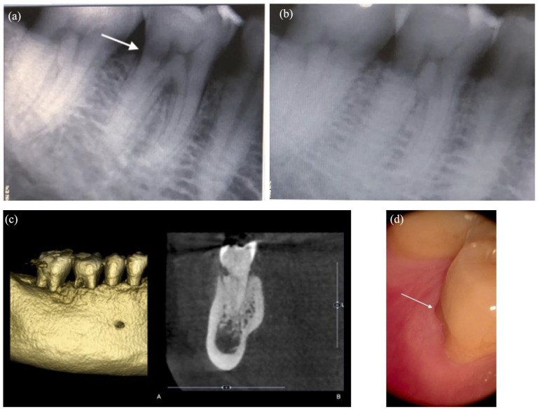
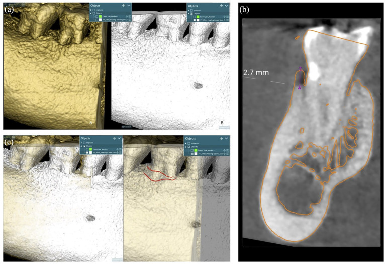
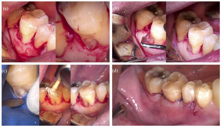
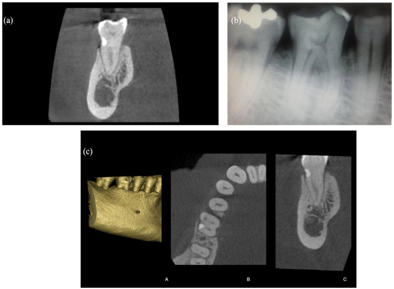
A 65-year-old Caucasian male was referred to an endodontic specialist practice in a private clinic in December 2019 for the management of an asymptomatic, radiolucent lesion located at the cervical level of the distal root of his right lower first molar, noticed during a routine periapical radiograph. After an accurate evaluation with cone-beam computed tomography (CBCT), the subgingival lesion was diagnosed as a supracrestal external cervical resorption (ECR), with a circumferential spread ⩽90°, confined to dentine without pulp involvement. The lesion was treated with the following sequence: (1) a full flap accessed the ECR, (2) the granulomatous tissue was removed from the root area, (3) the cavity was refreshed and filled with a well-refined and polished resin composite, (4) the flap was sutured at the cemento-enamel junction. A mandibular CBCT scan was performed before treatment, right after treatment, and 3 years postoperatively. Compared to the 3-year posttreatment CBCT scan, the immediate posttreatment one, revealed the absence of bone loss and an unexpected coronal bone remodeling with new bone formation over the treated lesion.

摘要

一名65岁的白种男性于2019年12月被转诊至一家私人诊所的牙髓病专科,以处理在常规根尖片检查时发现的位于其右下第一磨牙远中牙根颈部的无症状放射性透射区病变。经锥形束计算机断层扫描(CBCT)精确评估后,龈下病变被诊断为龈上型颈缘外吸收(ECR),圆周扩散≤90°,局限于牙本质且未累及牙髓。该病变按以下步骤进行治疗:(1)通过全层瓣暴露ECR;(2)从牙根区域清除肉芽组织;(3)清理窝洞并充填精心调制和抛光的树脂复合材料;(4)在牙骨质-釉质界处缝合瓣。在治疗前、治疗后即刻以及术后3年进行了下颌CBCT扫描。与术后3年的CBCT扫描相比,治疗后即刻的扫描显示无骨质丧失,且在治疗病变上方出现了意外的冠部骨重塑和新骨形成。

https://cdn.ncbi.nlm.nih.gov/pmc/blobs/9c2a/11320676/546175ecb665/10.1177_2050313X241269588-fig4.jpg
https://cdn.ncbi.nlm.nih.gov/pmc/blobs/9c2a/11320676/3be777cefdca/10.1177_2050313X241269588-fig1.jpg
https://cdn.ncbi.nlm.nih.gov/pmc/blobs/9c2a/11320676/b5b28ee00548/10.1177_2050313X241269588-fig2.jpg
https://cdn.ncbi.nlm.nih.gov/pmc/blobs/9c2a/11320676/733f8d80504e/10.1177_2050313X241269588-fig3.jpg
https://cdn.ncbi.nlm.nih.gov/pmc/blobs/9c2a/11320676/546175ecb665/10.1177_2050313X241269588-fig4.jpg

相似文献

1
Exceptional bone remodeling over composite restoration in the management of external root resorption: A case report.复合树脂修复治疗牙根外吸收中卓越的骨重塑:一例报告
SAGE Open Med Case Rep. 2024 Aug 12;12:2050313X241269588. doi: 10.1177/2050313X241269588. eCollection 2024.
2
Clinical and radiographic features of external cervical resorption - An observational study.临床和影像学特征的外部颈吸收-一项观察性研究。
Int Endod J. 2023 Dec;56(12):1475-1487. doi: 10.1111/iej.13968. Epub 2023 Oct 6.
3
Case report: pre-eruptive intra-coronal radiolucencies revisited.病例报告:再探萌出前牙冠内透射区
Eur Arch Paediatr Dent. 2012 Aug;13(4):221-6. doi: 10.1007/BF03262874.
4
Nonsurgical Management and 2-year Follow-up by means of Cone Beam Computed Tomography of an Invasive Cervical Resorption in a Molar.磨牙侵袭性颈部吸收的非手术治疗及通过锥形束计算机断层扫描进行的2年随访
J Contemp Dent Pract. 2018 Sep 1;19(9):1152-1156.
5
External cervical resorption: a three-dimensional classification.外部性颈吸收:一种三维分类法。
Int Endod J. 2018 Feb;51(2):206-214. doi: 10.1111/iej.12824. Epub 2017 Sep 13.
6
The Assessment and Management of External Cervical Resorption with Periapical Radiographs and Cone-beam Computed Tomography: A Clinical Study.根尖片和锥形束计算机断层扫描对外侧颈吸收的评估与处理:一项临床研究
J Endod. 2016 Oct;42(10):1435-40. doi: 10.1016/j.joen.2016.06.014. Epub 2016 Aug 6.
7
Prevalence and Characterization of External Cervical Resorption Using Cone Beam Computed Tomography.应用锥形束 CT 研究外部性宫颈吸收的发生情况和特征。
J Endod. 2024 Feb;50(2):164-172.e1. doi: 10.1016/j.joen.2023.11.003. Epub 2023 Nov 15.
8
Understanding external cervical resorption patterns in endodontically treated teeth.了解根管治疗后的牙齿的外部颈吸收模式。
Int Endod J. 2017 Dec;50(12):1116-1133. doi: 10.1111/iej.12744. Epub 2017 Feb 27.
9
External Cervical Resorption: A Case Report and Brief Review of the Literature, and Treatment Algorithms.外生型颈吸收:病例报告及文献复习,并提出治疗方案。
J Contemp Dent Pract. 2021 Mar 1;22(3):298-303.
10
Impact of Cone-beam Computed Tomography on Treatment Planning for External Cervical Resorption and a Novel Axial Slice-based Classification System.锥形束计算机断层扫描对外源性牙颈部吸收治疗计划的影响及一种新的基于轴向切片的分类系统。
J Endod. 2018 Feb;44(2):239-244. doi: 10.1016/j.joen.2017.10.001. Epub 2017 Dec 8.

本文引用的文献

1
Benefits of using magnification in dental specialties - a narrative review.牙科专业中使用放大技术的益处——一篇叙述性综述。
Med Pharm Rep. 2023 Jul;96(3):254-257. doi: 10.15386/mpr-2556. Epub 2023 Jul 27.
2
Repair of an Extensive External Cervical Resorption Lesion Using Intentional Replantation with Crown Rotation.采用带牙冠旋转的有意再植术修复广泛的颈外吸收病变
Case Rep Dent. 2023 Jun 26;2023:2103999. doi: 10.1155/2023/2103999. eCollection 2023.
3
Effect of sodium hypochlorite on bonding performance of universal adhesives to pulp chamber dentin.
次氯酸钠对通用型粘结剂与髓腔牙本质粘结性能的影响。
J Dent Sci. 2023 Jul;18(3):1116-1124. doi: 10.1016/j.jds.2022.11.007. Epub 2022 Nov 19.
4
Does endodontics influence radiological detection of external root resorption? an in vitro study.牙髓病学是否影响外部牙根吸收的放射学检测?一项体外研究。
BMC Oral Health. 2023 Apr 17;23(1):221. doi: 10.1186/s12903-023-02871-w.
5
[Pathogenesis and classification of tooth resorption].[牙齿吸收的发病机制与分类]
Zhonghua Kou Qiang Yi Xue Za Zhi. 2022 Nov 9;57(11):1177-1181. doi: 10.3760/cma.j.cn112144-20220513-00254.
6
Conservative Therapy of External Invasive Cervical Resorption with Adhesive Systems: A 6-Year Follow-Up Case Report and Literature Review.采用粘结系统对外部侵袭性颈部吸收进行保守治疗:一项6年随访病例报告及文献综述
Case Rep Dent. 2022 Oct 28;2022:9620629. doi: 10.1155/2022/9620629. eCollection 2022.
7
Risk factors for external root resorption of maxillary second molars associated with third molars.与第三磨牙相关的上颌第二磨牙牙根外吸收的危险因素。
Imaging Sci Dent. 2022 Sep;52(3):289-294. doi: 10.5624/isd.20220401. Epub 2022 May 13.
8
Tooth root resorption: A review.牙骨质吸收:综述。
Sci Prog. 2022 Jul-Sep;105(3):368504221109217. doi: 10.1177/00368504221109217.
9
External root resorption evaluated by CBCT 3D models superimposition.基于 CBCT 三维模型叠加评估的外部牙根吸收。
Dental Press J Orthod. 2022 Jun 10;27(2):e2219315. doi: 10.1590/2177-6709.27.2.e2219315.oar. eCollection 2022.
10
Endodontic management of a tooth with apical overfilling and perforating external root resorption: A case report.根尖超充并伴有外吸收穿孔牙齿的根管治疗:病例报告
Clin Case Rep. 2020 Oct 15;8(12):3278-3283. doi: 10.1002/ccr3.3406. eCollection 2020 Dec.