Allegheny General Hospital, 320 E North Ave, Pittsburgh, PA, 15201, USA.
Int Orthop. 2024 Oct;48(10):2743-2748. doi: 10.1007/s00264-024-06265-7. Epub 2024 Aug 15.
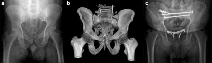
Fractures and dislocations of the pelvic ring are complex injuries that when treating require meticulous attention to detail and often specialized technical skill. These injuries can be the result of high-energy trauma, particularly in younger patients, or low energy trauma more often found in the elderly. Regardless of mechanism, these injuries lie on a spectrum of severity and can be treated conservatively or surgically. Percutaneous fixation under fluoroscopic guidance is the preferred standard technique when treating these fractures. This technique can be challenging for a variety of reasons including patient characteristics, intra-operative image quality, fracture morphology, among others.
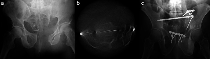
This retrospective study evaluated the use of intra-operative computed tomography (CT) using an O-arm imaging system for critical evaluation of fluoroscopic-guided screw placement in twenty-three patients. We retrospectively reviewed all cases of patients who were treated by three fellowship-trained orthopaedic traumatologists during a one-year span. Patients undergoing percutaneous pelvis fixation using both standard fluoroscopy and intraoperative CT with the Medtronic O-arm® (Minneapolis, MN) imaging system. Additionally, procedures performed included open reduction internal fixation (ORIF) of the pelvic ring, acetabulum, and associated extremity fractures.
Twenty-three patients were included in this study. On average, the use of intraoperative CT added 24.4 min in operative time. Five patients (21.7%) required implant adjustment after O-arm spin. Fourteen patients underwent additional post-operative CT. No secondary revision surgeries were attempted after any post-operative CT.
Our study suggests that intra-operative CT scan, compared to post-operative CT scan, can be utilized to prevent take-back surgery for misplaced implants and allow for adjustment in real-time.
骨盆环骨折和脱位是复杂的损伤,在治疗时需要细致入微地注意细节,并且通常需要专门的技术技能。这些损伤可能是高能创伤的结果,尤其是在年轻患者中,或者是低能量创伤更常见于老年患者。无论机制如何,这些损伤都存在严重程度的范围,可以保守治疗或手术治疗。在透视引导下经皮固定是治疗这些骨折的首选标准技术。由于各种原因,包括患者特征、术中图像质量、骨折形态等,该技术可能具有挑战性。
这项回顾性研究评估了在 23 名患者中使用术中计算机断层扫描(CT)通过 O 臂成像系统对透视引导螺钉放置进行关键评估的情况。我们回顾性地审查了在一年期间由三位 fellowship 培训的骨科创伤医生治疗的所有患者的所有病例。使用 Medtronic O-arm®(明尼阿波利斯,MN)成像系统进行标准透视和术中 CT 的患者接受了经皮骨盆固定术。此外,还进行了骨盆环、髋臼和相关肢体骨折的切开复位内固定(ORIF)手术。
本研究共纳入 23 例患者。平均而言,术中 CT 的使用增加了 24.4 分钟的手术时间。5 名患者(21.7%)在 O 臂旋转后需要调整植入物。14 名患者接受了额外的术后 CT。任何术后 CT 后均未尝试进行二次翻修手术。
与术后 CT 扫描相比,术中 CT 扫描可用于预防因植入物错位而进行的翻修手术,并允许实时调整。