Department of Neurology and Experimental Neurology, Charité-Universitätsmedizin Berlin, corporate member of Freie Universität and Humboldt-Universität zu Berlin, Berlin, Germany.
German Center for Neurodegenerative Diseases (DZNE) Berlin, Berlin, Germany.
Front Immunol. 2024 Aug 2;15:1404800. doi: 10.3389/fimmu.2024.1404800. eCollection 2024.
Patients suffering from neurological symptoms after COVID-19 vaccination (post-COVID-19 vaccination syndrome (PCVS)) have imposed an increasing challenge on medical practice, as diagnostic precision and therapeutic options are lacking. Underlying autoimmune dysfunctions, including autoantibodies, have been discussed in neurological disorders after SARS-CoV-2 infection and vaccination. Here, we describe the frequency and targets of autoantibodies against peripheral nervous system tissues in PCVS.
Sera from 50 PCVS patients with peripheral neurological symptoms after COVID-19 vaccination and 35 vaccinated healthy controls were used in this study. IgG autoreactivity was measured via indirect immunofluorescence assays on mouse sciatic nerve teased fibers. The frequencies of autoantibodies were compared between groups using Fisher's exact test. Serum anti-ganglioside antibodies were measured in ganglioside blots. Autoantibody target identification was performed using immunoprecipitation coupled to mass spectrometry. Subsequent target confirmation was conducted via cell-based assays and ELISA.
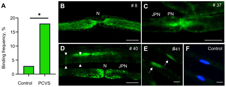
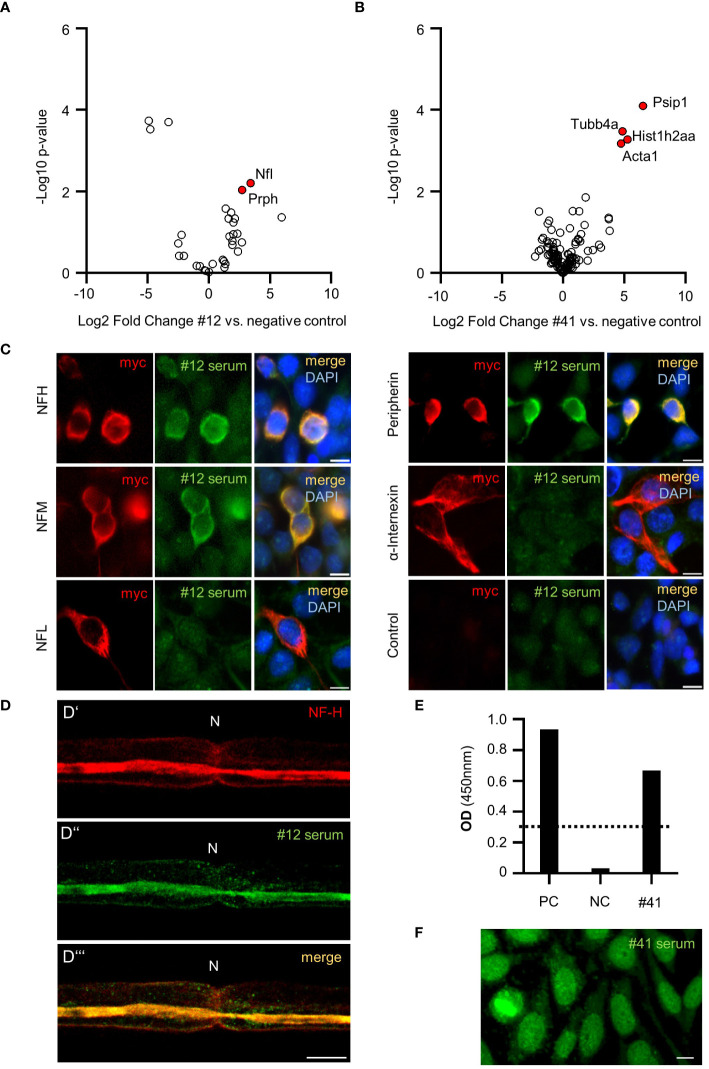
Compared with controls, PCVS patients had a significantly greater frequency of autoantibodies against peripheral nervous system structures (9/50(18%) vs 1/35(3%); p=0.04). Autoantibodies bound to paranodes (n=5), axons (n=4), Schmidt-Lanterman incisures (n=2) and Schwann cell nuclei (n=1). Conversely, antibodies against gangliosides were absent in PCVS patients. Target identification and subsequent confirmation revealed various subunits of neurofilaments as well as DFS-70 as autoantibody epitopes.
Our data suggest that autoantibodies against nervous system tissue could be relevant in PCVS patients. Autoantibodies against neurofilaments and cell nuclei with so far non-established links to this disease spectrum should be further elucidated to determine their biomarker potential.
患有 COVID-19 疫苗接种后出现神经系统症状的患者(COVID-19 疫苗接种后综合征 (PCVS))对医疗实践提出了越来越大的挑战,因为缺乏诊断精度和治疗选择。在 SARS-CoV-2 感染和接种疫苗后出现的神经障碍中,已经讨论了潜在的自身免疫功能障碍,包括自身抗体。在这里,我们描述了 PCVS 中针对周围神经系统组织的自身抗体的频率和靶标。
本研究使用了 50 例 COVID-19 疫苗接种后出现周围神经症状的 PCVS 患者和 35 例接种疫苗的健康对照者的血清。通过在小鼠坐骨神经 teased 纤维上进行间接免疫荧光测定来测量 IgG 自身反应性。使用 Fisher 精确检验比较组间的自身抗体频率。使用神经节苷脂印迹法测量血清抗神经节苷脂抗体。通过免疫沉淀结合质谱法进行自身抗体靶标鉴定。随后通过细胞测定和 ELISA 进行靶标确认。
与对照组相比,PCVS 患者外周神经系统结构的自身抗体频率明显更高(9/50(18%) vs 1/35(3%);p=0.04)。自身抗体结合到神经节旁区(n=5)、轴突(n=4)、施密特-兰坦曼切迹(n=2)和施万细胞核(n=1)。相反,PCVS 患者不存在针对神经节苷脂的抗体。靶标鉴定和随后的确认表明神经丝的各种亚单位以及 DFS-70 是自身抗体表位。
我们的数据表明,针对神经系统组织的自身抗体可能与 PCVS 患者有关。针对神经丝和细胞核的自身抗体与该疾病谱尚未建立的联系,应进一步阐明以确定其作为生物标志物的潜力。