• 文献检索
  • 文档翻译
  • 深度研究
  • 学术资讯
  • Suppr Zotero 插件Zotero 插件
  • 邀请有礼
  • 套餐&价格
  • 历史记录
应用&插件
Suppr Zotero 插件Zotero 插件浏览器插件Mac 客户端Windows 客户端微信小程序
定价
高级版会员购买积分包购买API积分包
服务
文献检索文档翻译深度研究API 文档MCP 服务
关于我们
关于 Suppr公司介绍联系我们用户协议隐私条款
关注我们

Suppr 超能文献

核心技术专利:CN118964589B侵权必究
粤ICP备2023148730 号-1Suppr @ 2026

文献检索

告别复杂PubMed语法,用中文像聊天一样搜索,搜遍4000万医学文献。AI智能推荐,让科研检索更轻松。

立即免费搜索

文件翻译

保留排版,准确专业,支持PDF/Word/PPT等文件格式,支持 12+语言互译。

免费翻译文档

深度研究

AI帮你快速写综述,25分钟生成高质量综述,智能提取关键信息,辅助科研写作。

立即免费体验

对比增强经颅多普勒和对比增强经胸超声心动图对卵圆孔未闭右向左分流的诊断价值:一项系统评价和荟萃分析。

The diagnostic value of contrast-enhanced transcranial Doppler and contrast-enhanced transthoracic echocardiography for right to left shunt in patent foramen ovale: a systematic review and meta-analysis.

作者信息

Zhang Dian, Jiang Li, Chen Yue-Nan, Pan Mei-Fang

机构信息

Department of Ultrasound, Xiangcheng People's Hospital, Suzhou, China.

出版信息

Front Neurol. 2024 Aug 2;15:1447964. doi: 10.3389/fneur.2024.1447964. eCollection 2024.

DOI:10.3389/fneur.2024.1447964
PMID:39157064
原文链接:https://pmc.ncbi.nlm.nih.gov/articles/PMC11327031/
Abstract

PURPOSE

To evaluate and compare the diagnostic value of contrast-enhanced transcranial Doppler (c-TCD) and contrast-enhanced transthoracic echocardiography (c-TTE) for right to left shunt (RLS) in patent foramen ovale (PFO) by meta-analysis.

METHODS

The literature included in the Cochrane Library, PubMed, and Embase were searched by using "contrast-enhanced transcranial Doppler (c-TCD), contrast-enhanced transthoracic echocardiography (c-TTE), patent foramen ovale (PFO), and right to left shunt (RLS)" as the keywords from inception through April 30, 2024. The diagnostic accuracy research quality assessment tool (QUADAS-2) was used to evaluate the quality of the included literature. The combined sensitivity, specificity, positive likelihood ratio (PLR), negative likelihood ratio (NLR), and Diagnostic odds ratio (DOR) were pooled, and a comprehensive ROC curve analysis was performed. Statistical software StataSE 12.0 and Meta-Disc 1.4 were used for data analysis.

RESULTS

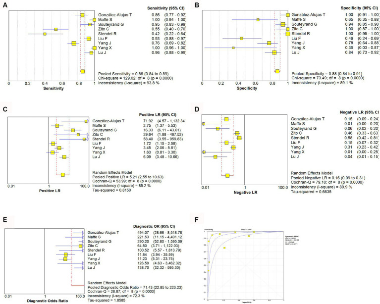
A total of 8,536 articles were retrieved, and 9 articles that met all inclusion criteria were included in this meta-analysis. The meta-analysis results show that the combined sensitivity, specificity, PLR, NLR, DOR, and area under the SROC curve of c-TCD for the diagnose of PFO-RLS were 0.91 (95% CI, 0.88-0.93), 0.87 (95% CI: 0.84-0.91), 6.0 (95% CI, 2.78-12.96), 0.10 (95% CI, 0.06-0.18), 91.61 (95% CI, 26.55-316.10), and 0.9681, respectively; the corresponding values of c-TTE were 0.86 (95% CI, 0.84-0.89), 0.88 (95% CI, 0.84-0.91), 5.21 (95% CI, 2.55-10.63), 0.16 (95% CI, 0.09-0.31), 71.43 (95% CI, 22.85-223.23), and 0.9532. The ROC curve shows that c-TCD has slightly higher diagnostic value for PFO than c-TTE, but there is no significant statistical difference (Z = 0.622, > 0.05). Deek funnel pattern showed no significant publication bias.

CONCLUSION

Both c-TCD and c-TTE have high diagnostic values for PFO-RLS. However, c-TCD has slightly higher sensitivity and lower specificity in diagnosing PFO-RLS compared to c-TTE. identifier [CRD42024544169].

摘要

目的

通过荟萃分析评估和比较对比增强经颅多普勒(c-TCD)和对比增强经胸超声心动图(c-TTE)对卵圆孔未闭(PFO)中右向左分流(RLS)的诊断价值。

方法

以“对比增强经颅多普勒(c-TCD)、对比增强经胸超声心动图(c-TTE)、卵圆孔未闭(PFO)、右向左分流(RLS)”为关键词,检索Cochrane图书馆、PubMed和Embase中从创刊至2024年4月30日的文献。采用诊断准确性研究质量评估工具(QUADAS-2)评估纳入文献的质量。汇总合并敏感度、特异度、阳性似然比(PLR)、阴性似然比(NLR)和诊断比值比(DOR),并进行全面的ROC曲线分析。使用统计软件StataSE 12.0和Meta-Disc 1.4进行数据分析。

结果

共检索到8536篇文章,9篇符合所有纳入标准的文章被纳入本荟萃分析。荟萃分析结果显示,c-TCD诊断PFO-RLS的合并敏感度、特异度、PLR、NLR、DOR及SROC曲线下面积分别为0.91(95%CI,0.88-0.93)、0.87(95%CI:0.84-0.9)、6.0(95%CI,2.78-12.96)、0.10(9%CI,0.06-0.18)、91.61(95%CI,26.55-316.10)和0.9681;c-TTE的相应值分别为0.86(95%CI,0.84-0.89)、0.88(95%CI,0.84-0.91)、5.21(95%CI,2.55-10.63)、0.16(95%CI,0.09-0.31)、71.43(95%CI,22.85-223.23)和0.9532。ROC曲线显示,c-TCD对PFO的诊断价值略高于c-TTE,但差异无统计学意义(Z=0.622,P>0.05)。Deek漏斗图显示无明显发表偏倚。

结论

c-TCD和c-TTE对PFO-RLS均具有较高的诊断价值。然而,与c-TTE相比,c-TCD在诊断PFO-RLS时具有略高的敏感度和较低的特异度。标识符[CRD42024544169]。

https://cdn.ncbi.nlm.nih.gov/pmc/blobs/28be/11327031/e9458e3b8f65/fneur-15-1447964-g004.jpg
https://cdn.ncbi.nlm.nih.gov/pmc/blobs/28be/11327031/48ef81b60b95/fneur-15-1447964-g001.jpg
https://cdn.ncbi.nlm.nih.gov/pmc/blobs/28be/11327031/4a5a7256969e/fneur-15-1447964-g002.jpg
https://cdn.ncbi.nlm.nih.gov/pmc/blobs/28be/11327031/93c70e083859/fneur-15-1447964-g003.jpg
https://cdn.ncbi.nlm.nih.gov/pmc/blobs/28be/11327031/e9458e3b8f65/fneur-15-1447964-g004.jpg

相似文献

1
The diagnostic value of contrast-enhanced transcranial Doppler and contrast-enhanced transthoracic echocardiography for right to left shunt in patent foramen ovale: a systematic review and meta-analysis.对比增强经颅多普勒和对比增强经胸超声心动图对卵圆孔未闭右向左分流的诊断价值:一项系统评价和荟萃分析。
Front Neurol. 2024 Aug 2;15:1447964. doi: 10.3389/fneur.2024.1447964. eCollection 2024.
2
Comparative diagnostic accuracy of transcranial Doppler and contrast-enhanced transthoracic echocardiography for the diagnosis of patent foramen ovale and atrial septal defect.经颅多普勒超声与增强经胸超声心动图诊断卵圆孔未闭与房间隔缺损的对比诊断准确性。
Panminerva Med. 2024 Jun;66(2):124-130. doi: 10.23736/S0031-0808.24.05123-1. Epub 2024 Apr 2.
3
Transcranial Doppler versus transthoracic echocardiography for the detection of patent foramen ovale in patients with cryptogenic cerebral ischemia: A systematic review and diagnostic test accuracy meta-analysis.经颅多普勒超声与经胸超声心动图在隐源性脑缺血患者卵圆孔未闭检测中的比较:系统评价和诊断试验准确性的荟萃分析。
Ann Neurol. 2016 Apr;79(4):625-35. doi: 10.1002/ana.24609. Epub 2016 Mar 11.
4
Comparative analysis of the diagnostic value of several methods for the diagnosis of patent foramen ovale.几种方法诊断卵圆孔未闭的诊断价值比较分析。
Echocardiography. 2021 May;38(5):790-797. doi: 10.1111/echo.15058. Epub 2021 Apr 18.
5
A Comparison of Transthroracic Echocardiograpy and Transcranial Doppler With Contrast Agent for Detection of Patent Foramen Ovale With or Without the Valsalva Maneuver.经胸超声心动图与经颅多普勒联合造影剂检测有无瓦尔萨尔瓦动作时卵圆孔未闭的比较
Medicine (Baltimore). 2015 Oct;94(43):e1937. doi: 10.1097/MD.0000000000001937.
6
Transthoracic second harmonic two- and three-dimensional echocardiography for detection of patent foramen ovale.经胸二次谐波二维及三维超声心动图用于检测卵圆孔未闭。
Eur J Echocardiogr. 2010 Jan;11(1):57-63. doi: 10.1093/ejechocard/jep165. Epub 2009 Nov 12.
7
Robot-Assisted Transcranial Doppler Versus Transthoracic Echocardiography for Right to Left Shunt Detection.机器人辅助经颅多普勒与经胸超声心动图用于检测右向左分流。
Stroke. 2023 Nov;54(11):2842-2850. doi: 10.1161/STROKEAHA.123.043380. Epub 2023 Oct 5.
8
PFO morphology for evaluation of c-TCD and c-TTE RLS grades.评估 c-TCD 和 c-TTE RLS 分级的卵圆孔未闭形态。
Eur J Med Res. 2022 Nov 3;27(1):228. doi: 10.1186/s40001-022-00855-0.
9
Accuracy of transcranial Doppler for the diagnosis of intracardiac right-to-left shunt: a bivariate meta-analysis of prospective studies.经颅多普勒超声诊断心内右向左分流的准确性:前瞻性研究的双变量荟萃分析。
JACC Cardiovasc Imaging. 2014 Mar;7(3):236-50. doi: 10.1016/j.jcmg.2013.12.011. Epub 2014 Feb 19.
10
Prevalence of patent foramen ovale in cryptogenic transient ischaemic attack and non-disabling stroke at older ages: a population-based study, systematic review, and meta-analysis.在年龄较大的隐源性短暂性脑缺血发作和非致残性卒中患者中卵圆孔未闭的患病率:基于人群的研究、系统评价和荟萃分析。
Lancet Neurol. 2018 Jul;17(7):609-617. doi: 10.1016/S1474-4422(18)30167-4. Epub 2018 Jun 7.

本文引用的文献

1
High-risk patent foramen ovale and elderly in cryptogenic stroke.隐匿性卒中中的高危卵圆孔未闭与老年人
J Stroke Cerebrovasc Dis. 2023 Nov;32(11):107344. doi: 10.1016/j.jstrokecerebrovasdis.2023.107344. Epub 2023 Sep 16.
2
Pearls and Pitfalls in the Transesophageal Echocardiographic Diagnosis of Patent Foramen Ovale.经食管超声心动图诊断卵圆孔未闭的要点与陷阱。
J Am Soc Echocardiogr. 2023 Sep;36(9):895-905.e3. doi: 10.1016/j.echo.2023.05.004. Epub 2023 May 15.
3
Multimodality imaging approach of patent foramen ovale: Practical considerations for transient ischemic attack/stroke.
卵圆孔未闭的多模态影像学检查方法:短暂性脑缺血发作/脑卒中的实用考虑因素。
J Clin Ultrasound. 2022 Oct;50(8):1166-1176. doi: 10.1002/jcu.23325.
4
Diagnostic Value of Micro-Bubble Transcranial Doppler Combined with Contrast Transthoracic Echocardiography in Cryptogenic Stroke Patients with Patent Foramen Ovale.微泡经颅多普勒超声联合经胸超声心动图造影对卵圆孔未闭伴隐源性脑卒中患者的诊断价值。
Neurol India. 2022 Jul-Aug;70(4):1403-1406. doi: 10.4103/0028-3886.355122.
5
Study of Transesophageal Echocardiography in Young Patients with Cryptogenic Stroke: Prevalence of Patent Foramen Ovale and Interpretation of the RoPE Score.经食管超声心动图在青年隐源性卒中患者中的研究:卵圆孔未闭的发生率及 RoPE 评分的解读。
Turk Kardiyol Dern Ars. 2022 Jul;50(5):314-319. doi: 10.5543/tkda.2022.21306.
6
Characteristics of Patent Foramen Ovale: Analysis from a Single Center.卵圆孔未闭的特征:来自单一中心的分析。
Cardiol Res Pract. 2022 Apr 6;2022:5430598. doi: 10.1155/2022/5430598. eCollection 2022.
7
Advances in the management of cardioembolic stroke associated with patent foramen ovale.卵圆孔未闭相关心源性脑梗死的管理进展。
BMJ. 2022 Feb 9;376:e063161. doi: 10.1136/bmj-2020-063161.
8
Persistent pathology of the patent foramen ovale: a review of the literature.持续性卵圆孔未闭的病理学:文献复习。
Med J Aust. 2021 Jul;215(2):89-93. doi: 10.5694/mja2.51141. Epub 2021 Jul 4.
9
Comparative analysis of the diagnostic value of several methods for the diagnosis of patent foramen ovale.几种方法诊断卵圆孔未闭的诊断价值比较分析。
Echocardiography. 2021 May;38(5):790-797. doi: 10.1111/echo.15058. Epub 2021 Apr 18.
10
The evolving role of echocardiography in the assessment of patent foramen ovale in patients with left-side thromboembolism.超声心动图在评估左侧血栓栓塞患者卵圆孔未闭中的作用演变。
Echocardiography. 2021 Apr;38(4):657-675. doi: 10.1111/echo.15018. Epub 2021 Mar 19.