Lu Cheng-Hsiu, Lian Wei-Shiung, Wu Re-Wen, Lin Yu-Han, Su Chia-Hao, Chen Chuan-Lin, Tai Ming-Hong, Chen Yu-Shan, Wang Shao-Yu, Chen Chao-Cheng, Wang Feng-Sheng
Department of Biomedical Imaging and Radiological Sciences, National Yang Ming Chiao Tung University, Taipei 112, Taiwan.
Institute of Biomedical Sciences, National Sun Yat-sen University, Kaohsiung 804, Taiwan.
APL Bioeng. 2024 Aug 19;8(3):036110. doi: 10.1063/5.0215273. eCollection 2024 Sep.
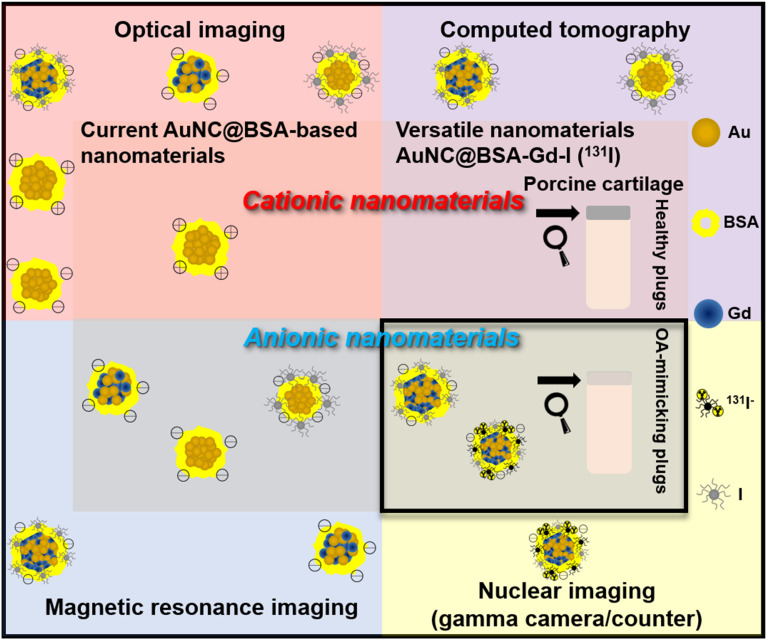
Cartilage damage, a common cause of osteoarthritis, requires medical imaging for accurate diagnosis of pathological changes. However, current instruments can acquire limited imaging information due to sensitivity and resolution issues. Therefore, multimodal imaging is considered an alternative strategy to provide valuable images and analyzes from different perspectives. Among all biomaterials, gold nanomaterials not only exhibit outstanding benefits as drug carriers, diagnostics, and radiosensitizers, but are also widely used as contrast agents, particularly for tumors. However, their potential for imaging cartilage damage is rarely discussed. In this study, we developed a versatile iodinated gadolinium-gold nanomaterial, AuNC@BSA-Gd-I, and its radiolabeled derivative, AuNC@BSA-Gd-I, for cartilage detection. With its small size, negative charge, and multimodal capacities, the probe can penetrate damaged cartilage and be detected or visualized by computed tomography, MRI, IVIS, and gamma counter. Additionally, the multimodal imaging potential of AuNC@BSA-Gd-I was compared to current multifunctional gold nanomaterials containing similar components, including anionic AuNC@BSA, AuNC@BSA-I, and AuNC@BSA-Gd as well as cationic AuNC@CBSA. Due to their high atomic numbers and fluorescent emission, AuNC@BSA nanomaterials could provide fundamental multifunctionality for imaging. By further modifying AuNC@BSA with additional imaging materials, their application could be extended to various types of medical imaging instruments. Nonetheless, our findings showed that each of the current nanomaterials exhibited excellent abilities for imaging cartilage with their predominant imaging modalities, but their versatility was not comparable to that of AuNC@BSA-Gd-I. Thus, AuNC@BSA-Gd-I could be served as a valuable tool in multimodal imaging strategies for cartilage assessment.
软骨损伤是骨关节炎的常见病因,需要医学成像来准确诊断病理变化。然而,由于灵敏度和分辨率问题,目前的仪器能够获取的成像信息有限。因此,多模态成像被认为是一种可从不同角度提供有价值图像和分析结果的替代策略。在所有生物材料中,金纳米材料不仅作为药物载体、诊断剂和放射增敏剂具有显著优势,还被广泛用作造影剂,尤其是用于肿瘤成像。然而,它们在软骨损伤成像方面的潜力却鲜有讨论。在本研究中,我们开发了一种多功能碘化钆金纳米材料AuNC@BSA-Gd-I及其放射性标记衍生物,用于软骨检测。该探针尺寸小、带负电荷且具有多模态能力,能够穿透受损软骨,并通过计算机断层扫描、磁共振成像、小动物活体成像系统和伽马计数器进行检测或可视化。此外,还将AuNC@BSA-Gd-I的多模态成像潜力与当前含有类似成分的多功能金纳米材料进行了比较,包括阴离子型AuNC@BSA、AuNC@BSA-I和AuNC@BSA-Gd以及阳离子型AuNC@CBSA。由于其高原子序数和荧光发射特性,AuNC@BSA纳米材料可为成像提供基本的多功能性。通过用其他成像材料对AuNC@BSA进行进一步修饰,其应用可扩展到各种类型的医学成像仪器。尽管如此,我们的研究结果表明,目前的每种纳米材料在其主要成像模式下对软骨成像均表现出优异的能力,但其多功能性无法与AuNC@BSA-Gd-I相媲美。因此,AuNC@BSA-Gd-I可作为软骨评估多模态成像策略中的一种有价值工具。