Collada-Carrasco Javier, Gómez-León Nieves, Castillo-Morales Valentina, Lumbreras-Fernández Blanca, Castañeda Santos, Rodríguez-Laval Víctor
Department of Radiology, Hospital Universitario de La Princesa, Autonomous University of Madrid, IIS-Princesa, Madrid, Spain.
Department of Nuclear Medicine, Hospital General Universitario Gregorio Marañón, Madrid, Spain.
Front Med (Lausanne). 2024 Aug 7;11:1432865. doi: 10.3389/fmed.2024.1432865. eCollection 2024.
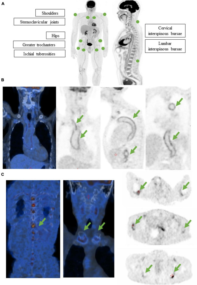
Large-vessel vasculitis (LVV) is a group of diseases characterized by inflammation of the aorta and its main branches, which includes giant cell arteritis (GCA), polymyalgia rheumatica (PMR), and Takayasu's arteritis (TAK). These conditions pose significant diagnostic and management challenges due to their diverse clinical presentations and potential for serious complications. F-fluorodeoxyglucose positron emission tomography-computed tomography (F-FDG-PET-CT) has emerged as a valuable imaging modality for the diagnosis and monitoring of LVV, offering insights into disease activity, extent, and response to treatment. F-FDG-PET-CT plays a crucial role in the diagnosis and management of LVV by allowing to visualize vessel involvement, assess disease activity, and guide treatment decisions. Studies have demonstrated the utility of F-FDG-PET-CT in distinguishing between LVV subtypes, evaluating disease distribution, and detecting extracranial involvement in patients with cranial GCA or PMR phenotypes. Additionally, F-FDG-PET-CT has shown promising utility in predicting clinical outcomes and assessing treatment response, based on the correlation between reductions in FDG uptake and improved disease control. Future research should focus on further refining PET-CT techniques, exploring their utility in monitoring treatment response, and investigating novel imaging modalities such as PET-MRI for enhanced diagnostic accuracy in LVV. Overall, F-FDG-PET-CT represents a valuable tool in the multidisciplinary management of LVV, facilitating timely diagnosis and personalized treatment strategies to improve patient outcomes.
大血管血管炎(LVV)是一组以主动脉及其主要分支炎症为特征的疾病,包括巨细胞动脉炎(GCA)、风湿性多肌痛(PMR)和大动脉炎(TAK)。由于这些疾病临床表现多样且有发生严重并发症的可能,因此在诊断和管理方面面临重大挑战。氟脱氧葡萄糖正电子发射断层扫描计算机断层扫描(F-FDG-PET-CT)已成为诊断和监测LVV的一种有价值的影像学检查方法,可提供有关疾病活动、范围及治疗反应的见解。F-FDG-PET-CT通过使血管受累情况可视化、评估疾病活动并指导治疗决策,在LVV的诊断和管理中发挥着关键作用。研究已证明F-FDG-PET-CT在区分LVV亚型、评估疾病分布以及检测具有颅部GCA或PMR表型患者的颅外受累情况方面的效用。此外,基于FDG摄取减少与疾病控制改善之间的相关性,F-FDG-PET-CT在预测临床结局和评估治疗反应方面也显示出有前景的效用。未来的研究应专注于进一步完善PET-CT技术,探索其在监测治疗反应方面的效用,并研究诸如PET-MRI等新型影像学检查方法,以提高LVV的诊断准确性。总体而言,F-FDG-PET-CT是LVV多学科管理中的一种有价值的工具,有助于及时诊断和制定个性化治疗策略以改善患者预后。