• 文献检索
  • 文档翻译
  • 深度研究
  • 学术资讯
  • Suppr Zotero 插件Zotero 插件
  • 邀请有礼
  • 套餐&价格
  • 历史记录
应用&插件
Suppr Zotero 插件Zotero 插件浏览器插件Mac 客户端Windows 客户端微信小程序
定价
高级版会员购买积分包购买API积分包
服务
文献检索文档翻译深度研究API 文档MCP 服务
关于我们
关于 Suppr公司介绍联系我们用户协议隐私条款
关注我们

Suppr 超能文献

核心技术专利:CN118964589B侵权必究
粤ICP备2023148730 号-1Suppr @ 2026

文献检索

告别复杂PubMed语法,用中文像聊天一样搜索,搜遍4000万医学文献。AI智能推荐,让科研检索更轻松。

立即免费搜索

文件翻译

保留排版,准确专业,支持PDF/Word/PPT等文件格式,支持 12+语言互译。

免费翻译文档

深度研究

AI帮你快速写综述,25分钟生成高质量综述,智能提取关键信息,辅助科研写作。

立即免费体验

通过光谱域光学相干断层扫描术测定的白内障手术顺利完成后3个月内视网膜各层厚度的变化。

Changes of individual retinal layer thickness post-uneventful cataract surgery determined by spectral-domain optical coherence tomography over a 3-months period.

作者信息

Großpötzl Manuel, Malle Eva Maria, Riedl Regina, Gran Jakob Daniel, Djavid Daniel, Posch-Pertl Laura, Glatz Wilfried Maximilian, Falb Thomas, Lindner Ewald, Wedrich Andreas, Ivastinovic Domagoj

机构信息

Department of Ophthalmology, Medical University of Graz, Graz, Austria.

Institute for Medical Informatics, Statistics and Documentation, Medical University of Graz, Austria.

出版信息

Heliyon. 2024 Jul 23;10(15):e35096. doi: 10.1016/j.heliyon.2024.e35096. eCollection 2024 Aug 15.

DOI:10.1016/j.heliyon.2024.e35096
PMID:39170147
原文链接:https://pmc.ncbi.nlm.nih.gov/articles/PMC11336489/
Abstract

PURPOSE

To evaluate retinal thickness changes of individual retinal layers using spectral-domain optical coherence tomography (SD-OCT) after uneventful cataract surgery over a 3-months period.

DESIGN

Prospective cohort study.

METHODS

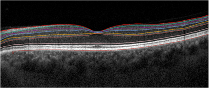
41 patients who underwent uneventful cataract surgery were included. Retinal SD-OCT images of both eyes were acquired preoperatively, 1 day after surgery as well as 1 month and 3 months postoperatively. Changes of retinal layer thickness were estimated after semi-automated segmentation for the following individual layers in the central subfield (CS, 1 mm) and inner ring (IR, 1-3 mm) of the early treatment diabetic retinopathy study (ETDRS) grid: retinal nerve fiber layer (RNFL), ganglion cell layer (GCL), inner plexiform layer (IPL), RNFL-GCL-IPL complex, inner nuclear layer (INL), outer plexiform layer (OPL), INL-OPL complex, outer nuclear layer (ONL), inner retina layer (IRL) and the total retina (TR). Furthermore, a sub-analysis with exclusion of patients devoid CME and an analysis in regard of patient age, lens status of the fellow eye, best corrected visual acuity and duration of surgery was conducted.

RESULTS

This study found significant alterations in all analysed retinal layers except for the RNFL (p = 0.33) and the GCL (p = 0.06) in the central subfield and the INL-OPL complex (p = 0.07) in the inner ring over the 3-months period (all p < 0.05). Retinal thickness decreases on the first postoperative day, followed by a significant increase 1 month after surgery and subsequent reduction at 3 months following uneventful cataract surgery could be observed.

CONCLUSION

These results assume the hypothesis that the apex of inflammatory response, characterized by an augmentation in the thickness of individual retinal layers, occurs around 1 month after uneventful cataract surgery, and subsequently experience a reduction in activity. Therefore, we suggest that additional therapy for cystoid macular edema does not have to be initiated as early as the first month in mild cases.

摘要

目的

使用光谱域光学相干断层扫描(SD-OCT)评估白内障手术顺利完成后3个月内各个视网膜层的视网膜厚度变化。

设计

前瞻性队列研究。

方法

纳入41例行白内障手术且过程顺利的患者。术前、术后1天以及术后1个月和3个月采集双眼的视网膜SD-OCT图像。在早期糖尿病视网膜病变研究(ETDRS)网格的中央子区域(CS,1mm)和内环(IR,1 - 3mm)中,对以下各个视网膜层进行半自动分割后估计视网膜层厚度的变化:视网膜神经纤维层(RNFL)、神经节细胞层(GCL)、内网状层(IPL)、RNFL - GCL - IPL复合体、内核层(INL)、外网状层(OPL)、INL - OPL复合体、外核层(ONL)、视网膜内层(IRL)和整个视网膜(TR)。此外,进行了一项排除无黄斑囊样水肿(CME)患者的亚分析,并针对患者年龄、对侧眼晶状体状态、最佳矫正视力和手术时长进行了分析。

结果

本研究发现,在3个月期间,除中央子区域的RNFL(p = 0.33)和GCL(p = 0.06)以及内环的INL - OPL复合体(p = 0.07)外,所有分析的视网膜层均有显著变化(所有p < 0.05)。可以观察到,白内障手术顺利完成后,术后第1天视网膜厚度下降,随后在术后1个月显著增加,3个月时又随后下降。

结论

这些结果支持这样的假设,即个体视网膜层厚度增加所表征的炎症反应高峰出现在白内障手术顺利完成后约1个月,随后炎症活动减弱。因此,我们建议在轻度病例中,不必早在第一个月就开始针对黄斑囊样水肿的额外治疗。

https://cdn.ncbi.nlm.nih.gov/pmc/blobs/5f21/11336489/4baeb4bad1a9/gr1.jpg

相似文献

1
Changes of individual retinal layer thickness post-uneventful cataract surgery determined by spectral-domain optical coherence tomography over a 3-months period.通过光谱域光学相干断层扫描术测定的白内障手术顺利完成后3个月内视网膜各层厚度的变化。
Heliyon. 2024 Jul 23;10(15):e35096. doi: 10.1016/j.heliyon.2024.e35096. eCollection 2024 Aug 15.
2
Comparison of Long-Term Automated Retinal Layer Segmentation Analysis of the Macula between Silicone Oil and Gas Tamponade after Vitrectomy for Rhegmatogenous Retinal Detachment.硅油和气体填充眼内手术后长期自动视网膜层分析黄斑比较。
Ophthalmic Res. 2020;63(6):524-532. doi: 10.1159/000506382. Epub 2020 Feb 10.
3
Mapping the thickness of retinal layers using Spectralis spectral domain optical coherence tomography in Indian eyes.利用 Spectralis 频域光学相干断层扫描仪对印度人眼视网膜各层厚度进行成像。
Indian J Ophthalmol. 2022 Aug;70(8):2990-2997. doi: 10.4103/ijo.IJO_81_22.
4
Anatomical effects of intravitreal anti-vascular endothelial growth factor injections on inner layers of the lesion-free retina.玻璃体腔抗血管内皮生长因子注射对无病变视网膜内层的解剖学影响。
Cutan Ocul Toxicol. 2021 Jun;40(2):135-139. doi: 10.1080/15569527.2021.1919136. Epub 2021 May 4.
5
Differential vulnerability of retinal layers to early age-related macular degeneration: evidence by SD-OCT segmentation analysis.视网膜各层对年龄相关性黄斑变性的易损性差异:SD-OCT 分割分析的证据。
Invest Ophthalmol Vis Sci. 2014 Jan 29;55(1):560-6. doi: 10.1167/iovs.13-12172.
6
Effects of different tamponade materials on macular segmentation after retinal detachment repair.不同眼内填充物对视网膜脱离修复术后黄斑区分割的影响。
Jpn J Ophthalmol. 2021 Mar;65(2):227-236. doi: 10.1007/s10384-020-00800-w. Epub 2021 Jan 9.
7
Optical coherence tomography analysis of inner and outer retinal layers in eyes with chiasmal compression caused by suprasellar tumours.鞍上肿瘤引起的视交叉受压眼的视网膜内层和外层的光相干断层扫描分析。
Acta Ophthalmol. 2020 May;98(3):e373-e380. doi: 10.1111/aos.14271. Epub 2019 Oct 10.
8
Evaluation of Structural Retinal Layer Alterations in Retinitis Pigmentosa.评估视网膜色素变性的结构视网膜层改变。
Rom J Ophthalmol. 2023 Oct-Dec;67(4):326-336. doi: 10.22336/rjo.2023.53.
9
Thickness mapping of individual retinal layers and sectors by Spectralis SD-OCT in Autosomal Dominant Optic Atrophy.Spectralis SD-OCT 对常染色体显性视神经萎缩个体视网膜各层和各象限的厚度进行测绘。
Acta Ophthalmol. 2018 May;96(3):251-256. doi: 10.1111/aos.13588. Epub 2017 Nov 1.
10
Effects of age, sex, and axial length on the three-dimensional profile of normal macular layer structures.年龄、性别和眼轴长度对正常黄斑层结构的三维形态的影响。
Invest Ophthalmol Vis Sci. 2011 Nov 11;52(12):8769-79. doi: 10.1167/iovs.11-8388.

引用本文的文献

1
The short term impact of uncomplicated cataract surgery on retinal layers thickness.单纯性白内障手术对视网膜各层厚度的短期影响。
Front Med (Lausanne). 2025 Jul 21;12:1537402. doi: 10.3389/fmed.2025.1537402. eCollection 2025.

本文引用的文献

1
The impact of surgeon's experience and sex on the incidence of cystoid macular edema after uneventful cataract surgery.手术医生经验和性别对无并发症白内障手术后囊样黄斑水肿发生率的影响。
PLoS One. 2022 Dec 27;17(12):e0279518. doi: 10.1371/journal.pone.0279518. eCollection 2022.
2
Comparison of the efficacy and safety of non-steroidal anti-inflammatory drugs and corticosteroid drugs for prevention of cystoid macular edema after cataract surgery.非甾体抗炎药与皮质类固醇药物预防白内障手术后黄斑囊样水肿的疗效及安全性比较。
Int Ophthalmol. 2023 Jan;43(1):271-284. doi: 10.1007/s10792-022-02426-y. Epub 2022 Sep 6.
3
Laser flare and cell photometry to measure inflammation after cataract surgery: a tool to predict the risk of cystoid macular edema.
激光散射细胞光度测定术在白内障术后炎症评估中的应用:预测黄斑囊样水肿风险的工具。
Int Ophthalmol. 2021 Jun;41(6):2293-2300. doi: 10.1007/s10792-021-01779-0. Epub 2021 Mar 21.
4
Preoperative central macular thickness as a risk factor for pseudophakic macular edema.术前中心黄斑厚度作为人工晶状体性黄斑水肿的一个危险因素。
Graefes Arch Clin Exp Ophthalmol. 2021 Jan;259(1):37-43. doi: 10.1007/s00417-020-04862-x. Epub 2020 Aug 11.
5
The Effects of Uncomplicated Cataract Surgery on Retinal Layer Thickness.单纯性白内障手术对视网膜层厚度的影响。
J Ophthalmol. 2018 Jun 5;2018:7218639. doi: 10.1155/2018/7218639. eCollection 2018.
6
Retinal layer segmentation in multiple sclerosis: a systematic review and meta-analysis.多发性硬化症中的视网膜层分割:系统评价和荟萃分析。
Lancet Neurol. 2017 Oct;16(10):797-812. doi: 10.1016/S1474-4422(17)30278-8. Epub 2017 Sep 12.
7
Changes of the Macular Ganglion Cell-Inner Plexiform Layer Thickness after Cataract Surgery in Glaucoma Patients.青光眼患者白内障手术后黄斑神经节细胞-内丛状层厚度的变化
J Ophthalmol. 2016;2016:9785939. doi: 10.1155/2016/9785939. Epub 2016 Dec 22.
8
Prevention of Cystoid Macular Edema After Cataract Surgery in Nondiabetic and Diabetic Patients: A Systematic Review and Meta-Analysis.非糖尿病和糖尿病患者白内障手术后囊样黄斑水肿的预防:一项系统评价和荟萃分析
Am J Ophthalmol. 2015 Nov;160(5):968-981.e33. doi: 10.1016/j.ajo.2015.07.032. Epub 2015 Jul 29.
9
Longitudinal analysis of the structural pattern of pseudophakic cystoid macular edema using multimodal imaging.使用多模态成像对人工晶状体眼黄斑囊样水肿的结构模式进行纵向分析。
Graefes Arch Clin Exp Ophthalmol. 2016 Jan;254(1):43-51. doi: 10.1007/s00417-015-3000-8. Epub 2015 Apr 12.
10
The effect of intracameral anesthesia on macular thickness and ganglion cell-inner plexiform layer thickness after uneventful phacoemulsification surgery: prospective and randomized controlled trial.眼内麻醉对无并发症超声乳化白内障吸除术后黄斑厚度和节细胞-内丛状层厚度的影响:前瞻性随机对照试验。
Graefes Arch Clin Exp Ophthalmol. 2014 Mar;252(3):433-9. doi: 10.1007/s00417-013-2557-3. Epub 2014 Jan 5.