Faculty of Dentistry, University of Toronto, Toronto, Ontario, Canada.
Division of Physical Medicine and Rehabilitation, Temerty Faculty of Medicine, University of Toronto, Toronto, Ontario, Canada.
PLoS One. 2024 Aug 22;19(8):e0307442. doi: 10.1371/journal.pone.0307442. eCollection 2024.
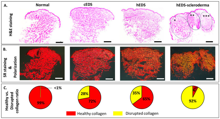
Ehlers-Danlos syndromes (EDS) represent a group of rare genetic disorders affecting connective tissues. Globally, approximately 1.5 million individuals suffer from EDS, with 10,000 reported cases in Canada alone. Understanding the histological properties of collagen in EDS has been challenging, but advanced techniques like atomic force microscopy (AFM) have opened up new possibilities for label-free skin imaging. This approach, which explores Type I collagen fibrils at the nanoscale, could potentially enhance EDS diagnosis and our knowledge of collagen type I-related connective tissue disorders. In the current study, we have employed AFM to examine ex-vivo skin biopsies from four individuals: one with classical EDS (cEDS), one with hypermobile EDS (hEDS), one with hEDS and Scleroderma (hEDS-Scleroderma), and one healthy control. Picrosirius red (PS) staining was used to highlight collagen differences in the samples. For each case, 14 images and 1400 force curves were obtained, with seven images and 700 force curves representing healthy collagen (PS-induced red staining) and the rest showcasing disrupted collagen (yellow staining). The results showed that PS staining was uniform throughout the control section, while cEDS and hEDS displayed localized areas of yellow staining. In the case of hEDS-Scleroderma, the yellow staining was widespread throughout the section. AFM images revealed irregular collagen fibrils in the disrupted, yellow-stained areas, contrasting with aligned and well-registered collagen fibrils in healthy, red-stained regions. Additionally, the study assessed the ability of non-AFM specialists to differentiate between healthy and disrupted collagen in AFM images, yielding substantial agreement among raters according to Fleiss's and Cohen's kappa scores (0.96 and 0.79±0.1, respectively). Biomechanical analysis revealed that normal healthy collagen exhibited a predominant population at 2.5 GPa. In contrast, EDS-affected collagen displayed subpopulations with lower compressive elastic modulus, indicating weaker collagen fibrils in EDS patients. Although these findings pertain to a limited number of cases, they offer valuable insights into the nanoscale collagen structure and biomechanics in individuals with EDS. Over time, these insights could be developed into specific biomarkers for the condition, improving diagnosis and treatment for EDS and related connective tissue disorders.
埃勒斯-当洛斯综合征(EDS)是一组罕见的遗传性疾病,影响结缔组织。全球约有 150 万人患有 EDS,仅在加拿大就报告了 10000 例。了解 EDS 中胶原蛋白的组织学特性一直具有挑战性,但原子力显微镜(AFM)等先进技术为无标记皮肤成像开辟了新的可能性。这种方法可以在纳米尺度上探索 I 型胶原纤维,有可能增强 EDS 的诊断和我们对 I 型胶原相关结缔组织疾病的认识。在目前的研究中,我们使用 AFM 检查了来自 4 个人的离体皮肤活检:1 个患有经典 EDS(cEDS),1 个患有高移动性 EDS(hEDS),1 个患有 hEDS 和硬皮病(hEDS-Scleroderma),1 个健康对照。苦味酸红(PS)染色用于突出样品中胶原蛋白的差异。对于每个病例,获得了 14 张图像和 1400 个力曲线,其中 7 张图像和 700 个力曲线代表健康的胶原蛋白(PS 诱导的红色染色),其余的则显示出破坏的胶原蛋白(黄色染色)。结果表明,PS 染色在对照部分均匀分布,而 cEDS 和 hEDS 显示出局部黄色染色区域。在 hEDS-Scleroderma 的情况下,黄色染色广泛分布在整个切片中。AFM 图像显示,在破坏的黄色染色区域中,胶原纤维不规则,与健康的红色染色区域中排列整齐且注册良好的胶原纤维形成对比。此外,该研究评估了非 AFM 专家在 AFM 图像中区分健康和破坏的胶原蛋白的能力,根据 Fleiss 和 Cohen 的kappa 评分(分别为 0.96 和 0.79±0.1),评分者之间存在显著的一致性。生物力学分析表明,正常健康的胶原蛋白在 2.5GPa 时表现出主要的种群。相比之下,EDS 影响的胶原蛋白显示出具有较低压缩弹性模量的亚种群,表明 EDS 患者的胶原纤维较弱。尽管这些发现仅涉及少数病例,但它们为 EDS 患者的纳米级胶原结构和生物力学提供了有价值的见解。随着时间的推移,这些见解可以发展为该病症的特定生物标志物,改善 EDS 和相关结缔组织疾病的诊断和治疗。