Mechatronics Research Lab, Massachusetts Institute of Technology, Cambridge, MA 02139, USA.
Biosensors (Basel). 2022 Dec 2;12(12):1116. doi: 10.3390/bios12121116.
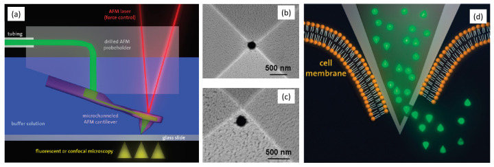
Visualization of biomedical samples in their native environments at the microscopic scale is crucial for studying fundamental principles and discovering biomedical systems with complex interaction. The study of dynamic biological processes requires a microscope system with multiple modalities, high spatial/temporal resolution, large imaging ranges, versatile imaging environments and ideally in-situ manipulation capabilities. Recent development of new Atomic Force Microscopy (AFM) capabilities has made it such a powerful tool for biological and biomedical research. This review introduces novel AFM functionalities including high-speed imaging for dynamic process visualization, mechanobiology with force spectroscopy, molecular species characterization, and AFM nano-manipulation. These capabilities enable many new possibilities for novel scientific research and allow scientists to observe and explore processes at the nanoscale like never before. Selected application examples from recent studies are provided to demonstrate the effectiveness of these AFM techniques.
在微观尺度下对生物医学样本进行原生环境下的可视化对于研究基本原理和发现具有复杂相互作用的生物医学系统至关重要。动态生物过程的研究需要具有多种模式、高空间/时间分辨率、大成像范围、多功能成像环境且理想情况下具有原位操作能力的显微镜系统。新型原子力显微镜(AFM)功能的最新发展使其成为生物学和生物医学研究的强大工具。本综述介绍了新型 AFM 功能,包括用于动态过程可视化的高速成像、具有力谱学的机械生物学、分子种类表征和 AFM 纳米操作。这些功能为新的科学研究提供了许多新的可能性,并使科学家能够以前所未有的方式观察和探索纳米尺度的过程。提供了来自最近研究的选定应用示例,以证明这些 AFM 技术的有效性。