Nougaret Stephanie, Lambregts Doenja M J, Beets Geerard L, Beets-Tan Regina G H, Blomqvist Lennart, Burling David, Denost Quentin, Gambacorta Maria A, Gui Benedetta, Klopp Ann, Lakhman Yulia, Maturen Kate E, Manfredi Riccardo, Petkovska Iva, Russo Luca, Shinagare Atul B, Stephenson James A, Tolan Damian, Venkatesan Aradhana M, Quyn Aaron J, Forstner Rosemarie
Department of Radiology, PINKCC lab, U1194, Montpellier Cancer Center, Montpellier, France.
Department of Radiology, Netherlands Cancer Institute, Amsterdam, The Netherlands.
Eur Radiol. 2025 May;35(5):2681-2691. doi: 10.1007/s00330-024-10940-z. Epub 2024 Aug 25.
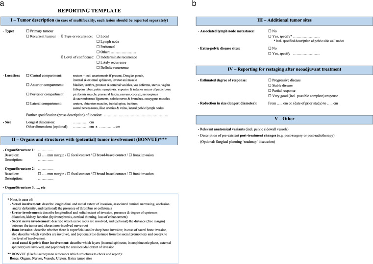
Pelvic exenteration (PE) is a radical surgical approach designed for the curative treatment of advanced pelvic malignancies, requiring en-bloc resection of multiple pelvic organs. While the procedure is radical, it has shown promise in enhancing long-term survival and is now comparable in surgical mortality to elective resections for primary pelvic cancers. Imaging plays a crucial role in preoperative planning, with MRI, CT, and PET/CT being pivotal in assessing the extent of cancer and formulating a surgical roadmap. This paper presents clinical practice guidelines for imaging in the context of PE, developed jointly by ESGAR, SAR, ESUR, and the PelvEx Collaborative. These guidelines aim to standardize imaging protocols and reporting to improve the preoperative assessment and facilitate decision-making in the multidisciplinary treatment of pelvic cancers. Our recommendations underscore the importance of a multidisciplinary approach and the need for clear and precise imaging reports to optimize patient care. CLINICAL RELEVANCE STATEMENT: Our recommendations underscore the importance of a multidisciplinary approach and the need for clear and precise imaging reports to optimize patient care. KEY POINTS: MRI is mandatory for local staging in pelvic exenteration. Structured reporting (using the template provided in this guide) is recommended. Multidisciplinary review of imaging is critical for surgical planning.
盆腔脏器清除术(PE)是一种根治性手术方法,旨在治疗晚期盆腔恶性肿瘤,需要整块切除多个盆腔器官。虽然该手术是根治性的,但已显示出提高长期生存率的前景,目前其手术死亡率与原发性盆腔癌的择期切除术相当。影像学在术前规划中起着关键作用,MRI、CT和PET/CT对于评估癌症范围和制定手术路线图至关重要。本文介绍了由欧洲胃肠道和腹部放射学会(ESGAR)、西班牙放射学会(SAR)、欧洲泌尿生殖放射学会(ESUR)和盆腔脏器清除协作组联合制定的PE影像学临床实践指南。这些指南旨在规范影像学检查方案和报告,以改善术前评估并促进盆腔癌多学科治疗中的决策制定。我们的建议强调了多学科方法的重要性以及清晰准确的影像学报告对于优化患者护理的必要性。临床相关性声明:我们的建议强调了多学科方法的重要性以及清晰准确的影像学报告对于优化患者护理的必要性。关键点:MRI对于盆腔脏器清除术的局部分期是必需的。建议采用结构化报告(使用本指南提供的模板)。影像学的多学科评估对于手术规划至关重要。