Eckstein Felix, Walter-Rittel Thula Cannon, Chaudhari Akshay S, Brisson Nicholas M, Maleitzke Tazio, Duda Georg N, Wisser Anna, Wirth Wolfgang, Winkler Tobias
Research Program for Musculoskeletal Imaging, Center for Anatomy & Cell Biology, Paracelsus Medical University, Salzburg, Austria.
Ludwig Boltzmann Institute for Arthritis and Rehabilitation (LBIAR), Paracelsus Medical University, Salzburg, Austria.
Osteoarthr Cartil Open. 2024 Jul 23;6(3):100505. doi: 10.1016/j.ocarto.2024.100505. eCollection 2024 Sep.
This expert opinion paper proposes a design for a state-of-the-art magnetic resonance image (MRI) acquisition protocol for knee osteoarthritis clinical trials in early and advanced disease. Semi-quantitative and quantitative imaging endpoints are supported, partly amendable to automated analysis. Several (peri-) articular tissues and pathologies are covered, including synovitis.
A PubMed literature search was conducted, with focus on the past 5 years. Further, osteoarthritis imaging experts provided input. Specific MRI sequences, orientations, spatial resolutions and parameter settings were identified to align with study goals. We strived for implementation on standard clinical scanner hardware, with a net acquisition time ≤30 min.
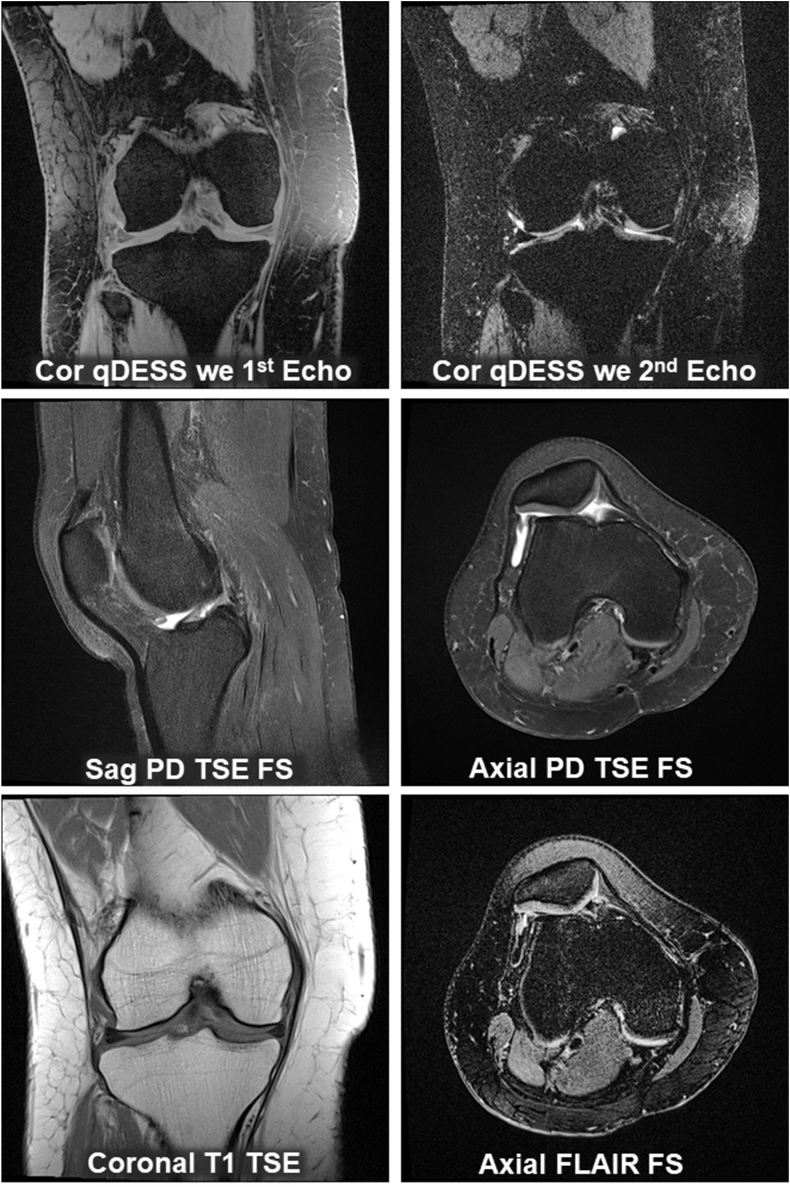
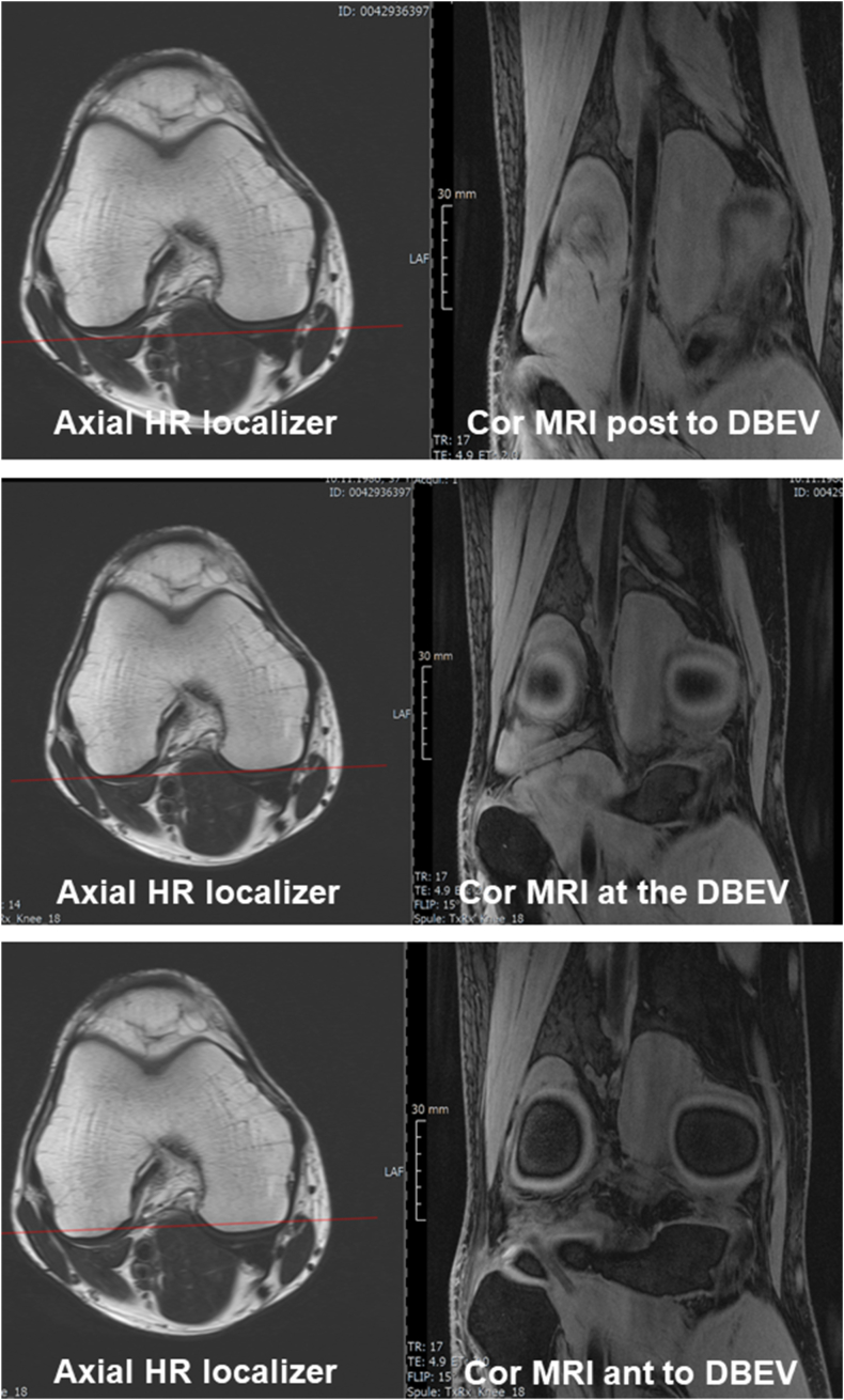
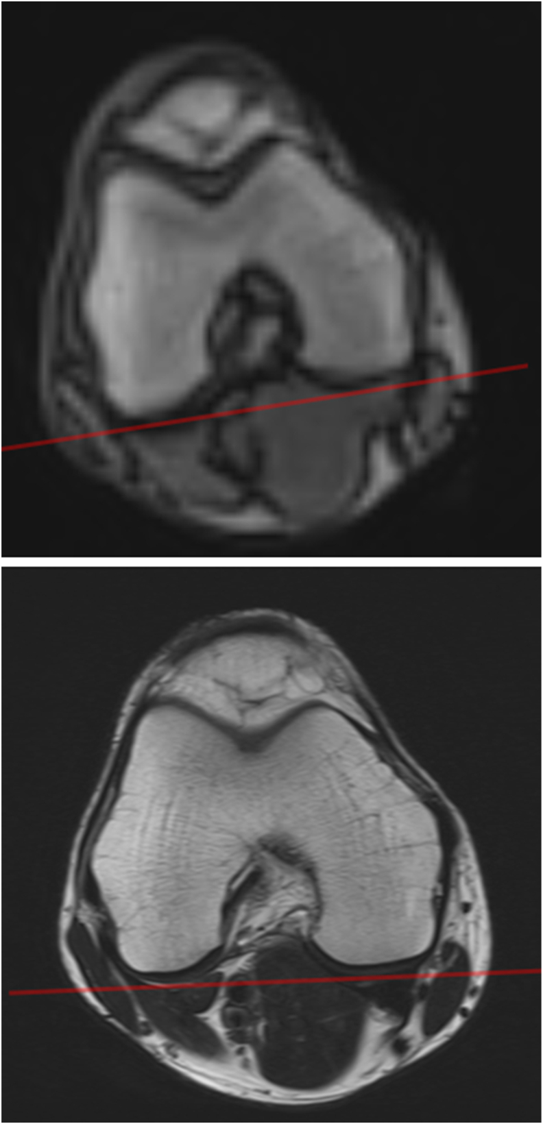
Short- and long-term longitudinal MRIs should be obtained at ≥1.5T, if possible without hardware changes during the study. We suggest a series of gradient- and spin-echo-sequences, supporting MOAKS, quantitative analysis of cartilage morphology and T2, and non-contrast-enhanced depiction of synovitis. These sequences should be properly aligned and positioned using localizer images. One of the sequences may be repeated in each participant (re-test), optimally at baseline and follow-up, to estimate within-study precision. All images should be checked for quality and protocol-adherence as soon as possible after acquisition. Alternative approaches are suggested that expand on the structural endpoints presented.
We aim to bridge the gap between technical MRI acquisition guides and the wealth of imaging literature, proposing a balance between image acquisition efficiency (time), safety, and technical/methodological diversity. This approach may entertain scientific innovation on tissue structure and composition assessment in clinical trials on disease modification of knee osteoarthritis.
本专家意见论文针对早期和晚期疾病的膝关节骨关节炎临床试验,提出了一种最先进的磁共振成像(MRI)采集方案设计。该方案支持半定量和定量成像终点,部分可用于自动分析。涵盖了几种(关节周围)组织和病理情况,包括滑膜炎。
进行了PubMed文献检索,重点关注过去5年的文献。此外,骨关节炎成像专家提供了意见。确定了特定的MRI序列、方位、空间分辨率和参数设置,以符合研究目标。我们力求在标准临床扫描仪硬件上实施,净采集时间≤30分钟。
如果可能,应在≥1.5T的磁场强度下获取短期和长期纵向MRI,且在研究期间无需更换硬件。我们建议采用一系列梯度回波和自旋回波序列,以支持MOAKS、软骨形态和T2的定量分析以及滑膜炎的非增强成像。这些序列应使用定位图像进行正确对齐和定位。其中一个序列可在每个参与者中重复(重新测试),最佳时间为基线和随访时,以评估研究内的精度。采集后应尽快检查所有图像的质量和是否符合方案要求。还提出了替代方法,以扩展所呈现的结构终点。
我们旨在弥合MRI技术采集指南与丰富的成像文献之间的差距,在图像采集效率(时间)、安全性和技术/方法多样性之间取得平衡。这种方法可能会促进膝关节骨关节炎疾病修饰临床试验中组织结构和成分评估的科学创新。