Liu Ming-Ming, Liang Yu-Ting, Jin Er-Hu
Department of Radiology, Beijing Friendship Hospital, Capital Medical University, Beijing 100050, China.
Department of Radiology, Beijing Obstetrics and Gynecology Hospital, Capital Medical University, Beijing Maternal and Child Health Care Hospital, Beijing 100006, China.
World J Clin Cases. 2024 Aug 26;12(24):5583-5588. doi: 10.12998/wjcc.v12.i24.5583.
Endometrial cancer is a kind of well-known tumors of female genitourinary system. Cervical stromal invasion is an adverse factor for poor prognosis of endometrial cancer. There is still controversy regarding the use of magnetic resonance imaging (MRI) in the diagnosis of cervical stromal invasion of endometrial cancer. The diagnosis of cervical stromal invasion varies significantly between different observers and institutions. We present a limited case series of the particular pattern of endometrial cancer, which infiltrates the cervical stroma and is often overlooked.
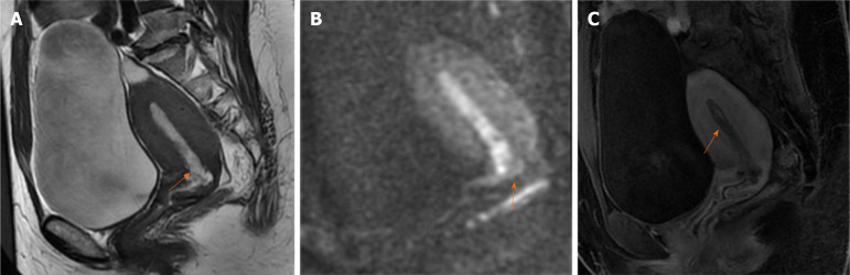
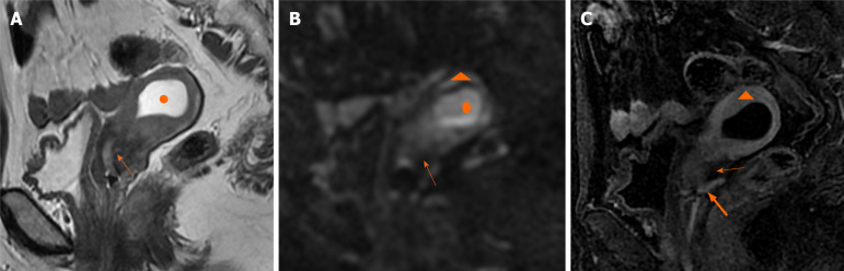
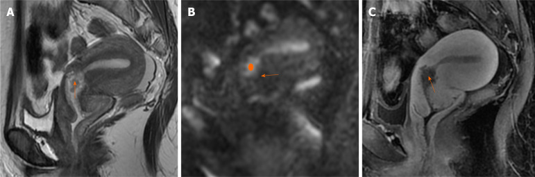
We present three cases of endometrial carcinoma with cervical stromal invasion with cancer-free uterine cavity. One patient, a reproductive-aged woman, exhibited irregular menstruation and was diagnosed with endometrial polyps by hysteroscopy and segmental curettage. A MRI scan revealed polypoid nodules within the internal cervical orifice. The other two cases were postmenopausal women who presented with abnormal vaginal bleeding. Hysteroscopy and segmental curettage suggested atypical hyperplasia of the endometrium. MRI scans did not detect any malignant signs in the endometrium. In one case, a non-thickened endometrium was observed, while in another, hyperplasia of the endometrium was seen. Notably, none of these patients had malignant tumors identified in the uterine cavity MRI scans. However, postoperative pathological results following hysterectomy consistently indicated cervical stromal invasion.
Cervical stromal invasion is easily missed if no cancer is found in the uterine body on MRI. Immunohistochemistry of endoscopic curettage specimens should be conducted to avoid underestimation of the disease.
子宫内膜癌是女性生殖泌尿系统一种常见的肿瘤。宫颈间质浸润是子宫内膜癌预后不良的一个不利因素。在子宫内膜癌宫颈间质浸润的诊断中,磁共振成像(MRI)的应用仍存在争议。不同观察者和机构对宫颈间质浸润的诊断差异很大。我们报告了一系列有限的特殊类型子宫内膜癌病例,这类癌症浸润宫颈间质,常被忽视。
我们报告了3例宫颈间质浸润且宫腔无癌的子宫内膜癌病例。1例育龄期女性患者,月经不规律,经宫腔镜检查和分段刮宫诊断为子宫内膜息肉。MRI扫描显示宫颈内口有息肉样结节。另外2例为绝经后女性,表现为异常阴道出血。宫腔镜检查和分段刮宫提示子宫内膜非典型增生。MRI扫描未在子宫内膜中检测到任何恶性征象。1例观察到子宫内膜未增厚,另1例可见子宫内膜增生。值得注意的是,这些患者的MRI扫描均未在宫腔内发现恶性肿瘤。然而,子宫切除术后的病理结果均显示存在宫颈间质浸润。
如果MRI检查未在子宫体发现癌症,宫颈间质浸润很容易被漏诊。应进行内镜刮宫标本的免疫组化检查,以避免对疾病的低估。