Bi Qiu, Bi Guoli, Wang Junna, Zhang Jie, Li Hongliang, Gong Xiarong, Ren Lixiang, Wu Kunhua
Department of MRI, the First People' s Hospital of Yunnan Province, the Affiliated Hospital of Kunming University of Science and Technology, No. 157 Jinbi Road, Kunming 650032, Yunnan, China.
Center of Infectious Diseases, West China Hospital of Sichuan University, No. 37 Guoxue Lane, Wuhou District, Chengdu 610041, Sichuan, China.
J Cancer. 2021 Jan 1;12(3):754-764. doi: 10.7150/jca.52797. eCollection 2021.
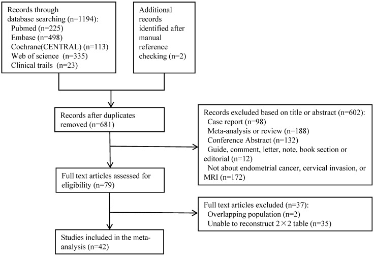
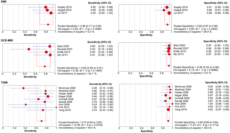
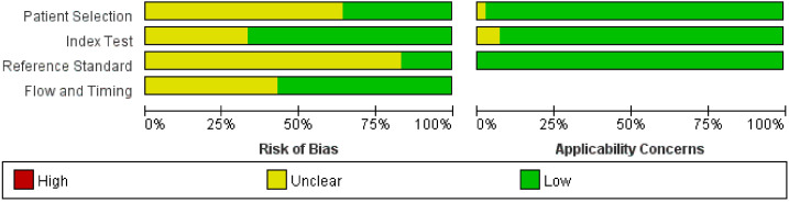
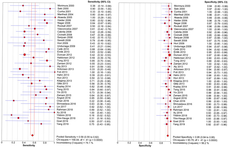
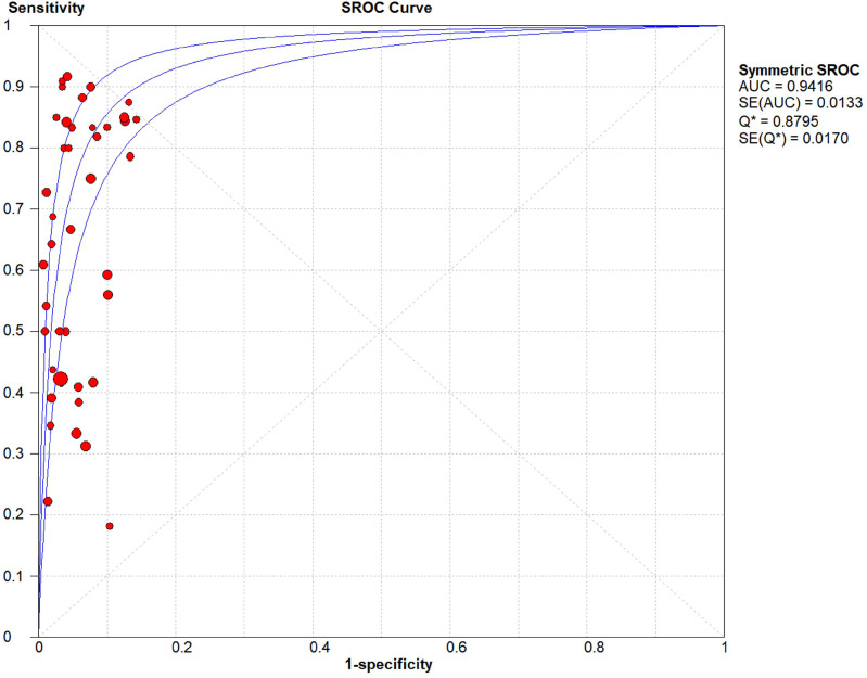
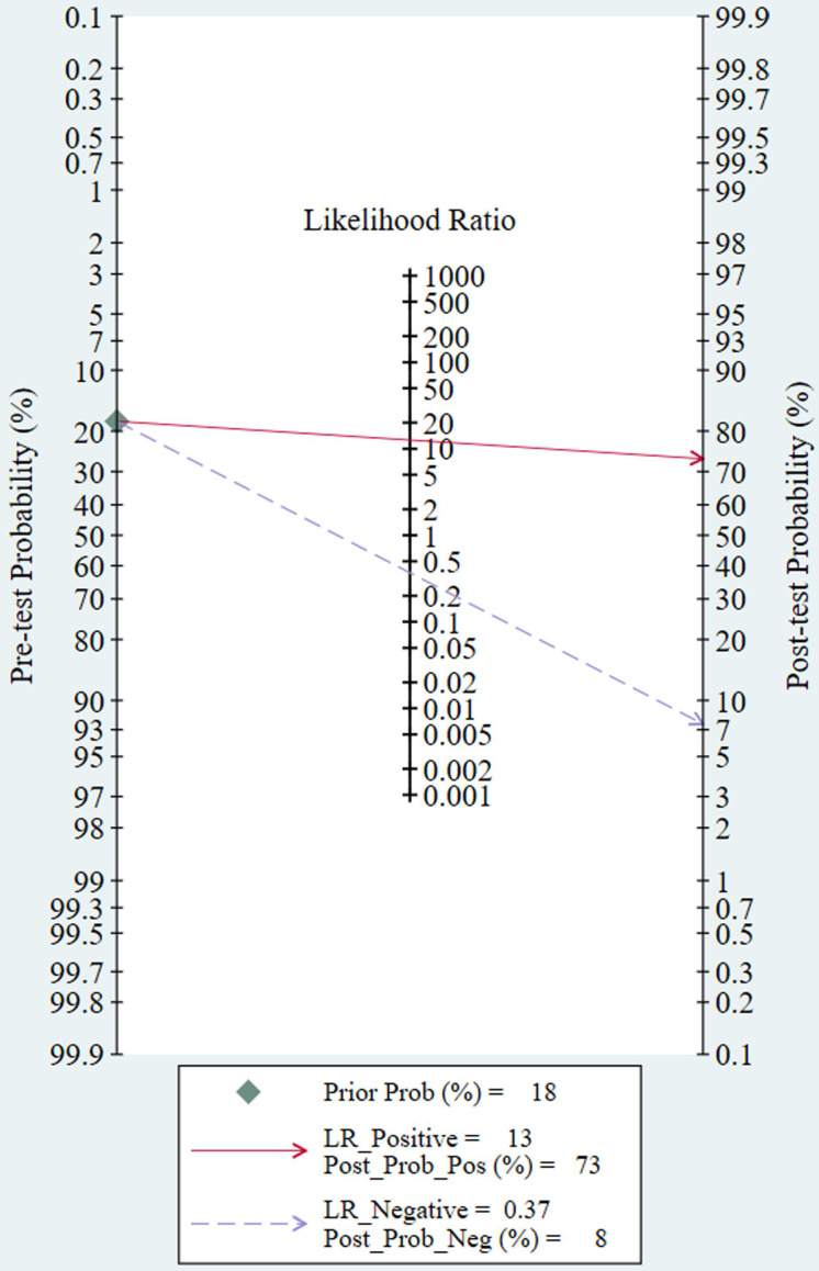
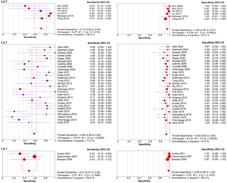
To evaluate the diagnostic accuracy of magnetic resonance imaging (MRI) in the preoperative assessment of cervical invasion and to analyse the influence of different imaging protocols in patients with endometrial carcinoma. An extensive search of articles about MRI for assessing cervical invasion in patients with endometrial carcinoma was performed on PubMed, Embase, Web of Science, Cochrane Library, and Clinical Trials from January 2000 to July 2020. Two reviewers independently evaluated the methodological quality of each study by using the Quality Assessment of Diagnostic Accuracy Studies-2 (QUADAS-2). Diagnostic accuracy results and additional useful information were extracted. The pooled estimation data was obtained by statistical analysis. A total of 42 eligible studies were included in the meta-analysis. Significant evidence of heterogeneity was found for detecting cervical invasion ( = 74.1%, = 0.00 for sensitivity and = 56.2%, = 0.00 for specificity). The pooled sensitivity and specificity of MRI were 0.58 and 0.95 respectively. The use of higher field strength (3.0 T) demonstrated higher pooled sensitivity (0.74). Using diffusion weighted imaging (DWI) alone presented higher pooled sensitivity (0.86) than using other sequences. The studies that used dynamic contrast-enhanced MRI (DCE-MRI) alone showed higher sensitivity (0.80) and specificity (0.96) than those that used T2-weighted imaging (T2WI) alone. MRI shows high specificity for detecting cervical infiltration in endometrial carcinoma. Using DWI or a 3.0-T device may improve the pooled sensitivity. DCE-MRI demonstrates higher pooled sensitivity and specificity than T2WI.
评估磁共振成像(MRI)在子宫内膜癌术前宫颈浸润评估中的诊断准确性,并分析不同成像方案对子宫内膜癌患者的影响。对2000年1月至2020年7月期间在PubMed、Embase、Web of Science、Cochrane图书馆和临床试验中关于MRI评估子宫内膜癌患者宫颈浸润的文章进行了广泛检索。两名 reviewers 使用诊断准确性研究质量评估-2(QUADAS-2)独立评估每项研究的方法学质量。提取诊断准确性结果和其他有用信息。通过统计分析获得汇总估计数据。荟萃分析共纳入42项符合条件的研究。在检测宫颈浸润方面发现了显著的异质性证据(敏感性 = 74.1%,I² = 0.00;特异性 = 56.2%,I² = 0.00)。MRI 的汇总敏感性和特异性分别为0.58和0.95。使用更高场强(3.0 T)显示出更高的汇总敏感性(0.74)。单独使用扩散加权成像(DWI)的汇总敏感性(0.86)高于使用其他序列。单独使用动态对比增强MRI(DCE-MRI)的研究比单独使用T2加权成像(T2WI)的研究显示出更高的敏感性(0.80)和特异性(0.96)。MRI对检测子宫内膜癌宫颈浸润具有高特异性。使用DWI或3.0-T设备可能会提高汇总敏感性。DCE-MRI的汇总敏感性和特异性高于T2WI。