• 文献检索
  • 文档翻译
  • 深度研究
  • 学术资讯
  • Suppr Zotero 插件Zotero 插件
  • 邀请有礼
  • 套餐&价格
  • 历史记录
应用&插件
Suppr Zotero 插件Zotero 插件浏览器插件Mac 客户端Windows 客户端微信小程序
定价
高级版会员购买积分包购买API积分包
服务
文献检索文档翻译深度研究API 文档MCP 服务
关于我们
关于 Suppr公司介绍联系我们用户协议隐私条款
关注我们

Suppr 超能文献

核心技术专利:CN118964589B侵权必究
粤ICP备2023148730 号-1Suppr @ 2026

文献检索

告别复杂PubMed语法,用中文像聊天一样搜索,搜遍4000万医学文献。AI智能推荐,让科研检索更轻松。

立即免费搜索

文件翻译

保留排版,准确专业,支持PDF/Word/PPT等文件格式,支持 12+语言互译。

免费翻译文档

深度研究

AI帮你快速写综述,25分钟生成高质量综述,智能提取关键信息,辅助科研写作。

立即免费体验

基于氟代脱氧葡萄糖正电子发射断层扫描/计算机断层扫描(F-FDG PET/CT)及磁共振成像(MRI)的子宫颈鳞状细胞癌阴蒂转移

Clitoral metastasis from squamous cell carcinoma of the cervix on F-FDG PET/CT and MRI.

作者信息

Vu Ryan, Tseng Jill, Vu Peter, Stelling Adam

机构信息

Stanford University.

University of California in Irvine.

出版信息

Radiol Case Rep. 2024 Aug 2;19(10):4508-4512. doi: 10.1016/j.radcr.2024.06.091. eCollection 2024 Oct.

DOI:10.1016/j.radcr.2024.06.091
PMID:39188631
原文链接:https://pmc.ncbi.nlm.nih.gov/articles/PMC11345129/
Abstract

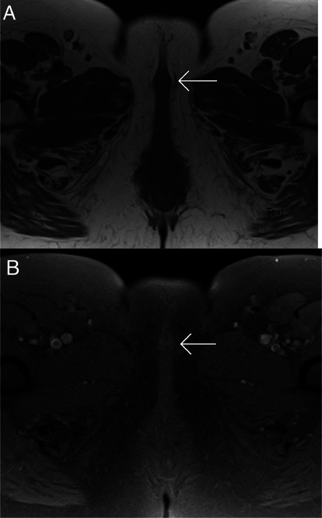
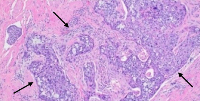
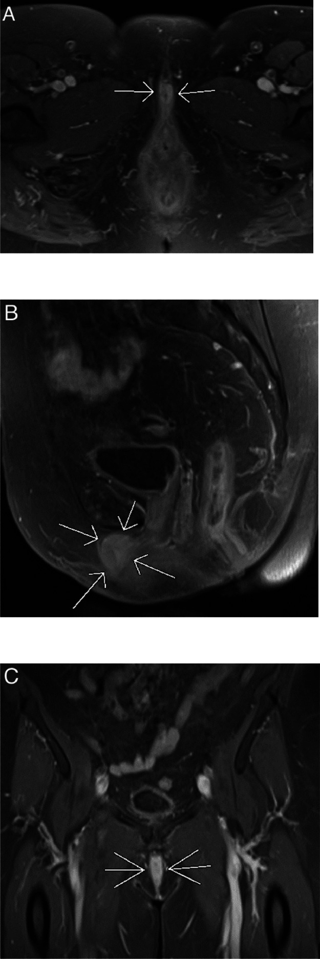
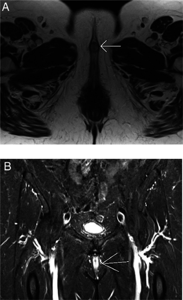
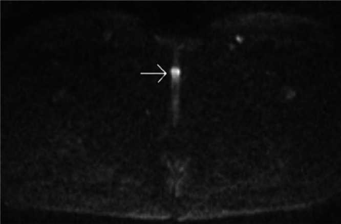
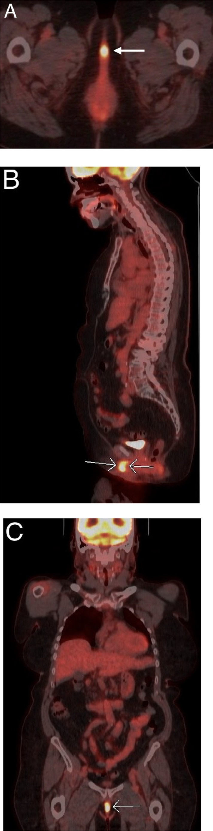
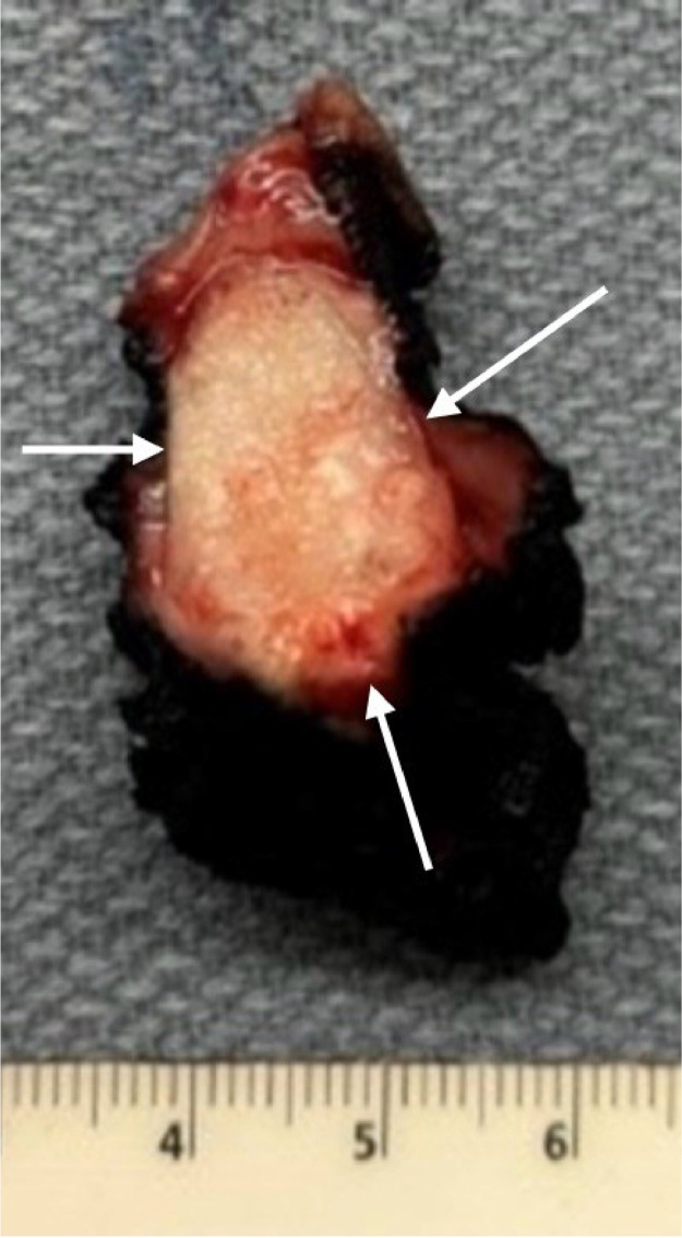
Metastasis to the clitoris is extremely rare. Here, we report a highly unusual case of high-grade squamous cell carcinoma of the cervix metastasizing to the clitoris a year following surgery and chemoradiotherapy. The patient presented with a painless clitoral mass identified through physical examination. Magnetic resonance imaging (MRI) showed a diffusely enhancing clitoral mass with hyperintense signals on diffusion-weighted imaging (DWI) and fluid-sensitive T2-weighted (T2W) sequences. This malignant tumor was detected by fluorine-fluorodeoxyglucose positron emission tomography/computed tomography (F-FDG PET/CT) due to its high FDG uptake. Pathological examination confirmed clitoral metastasis. Clitoral metastasis, although exceedingly rare, should be considered in cervical cancer patients presenting with clitoral masses on physical examination and imaging, particularly in those with advanced stages. Our case report is unique because it represents a recurrence in a patient initially diagnosed with early-stage cancer.

摘要

阴蒂转移极为罕见。在此,我们报告一例极为特殊的病例,一名宫颈高级别鳞状细胞癌患者在手术及放化疗一年后出现阴蒂转移。患者经体格检查发现有无痛性阴蒂肿物。磁共振成像(MRI)显示阴蒂肿物弥漫性强化,在扩散加权成像(DWI)及液体敏感的T2加权(T2W)序列上呈高信号。由于该恶性肿瘤对氟脱氧葡萄糖摄取较高,通过氟-氟脱氧葡萄糖正电子发射断层扫描/计算机断层扫描(F-FDG PET/CT)检测到了它。病理检查证实为阴蒂转移。阴蒂转移虽极为罕见,但对于体格检查及影像学检查发现有阴蒂肿物的宫颈癌患者,尤其是晚期患者,应予以考虑。我们的病例报告很独特,因为它代表了一名最初诊断为早期癌症患者的复发情况。

https://cdn.ncbi.nlm.nih.gov/pmc/blobs/8704/11345129/2fb84af9264a/gr7.jpg
https://cdn.ncbi.nlm.nih.gov/pmc/blobs/8704/11345129/b4e6a19c565f/gr1.jpg
https://cdn.ncbi.nlm.nih.gov/pmc/blobs/8704/11345129/8f2b1b408d67/gr2.jpg
https://cdn.ncbi.nlm.nih.gov/pmc/blobs/8704/11345129/76b8f2c40565/gr3.jpg
https://cdn.ncbi.nlm.nih.gov/pmc/blobs/8704/11345129/49dd7bd71ad3/gr4.jpg
https://cdn.ncbi.nlm.nih.gov/pmc/blobs/8704/11345129/9a6b4b257b64/gr5.jpg
https://cdn.ncbi.nlm.nih.gov/pmc/blobs/8704/11345129/76ee5358db6f/gr6.jpg
https://cdn.ncbi.nlm.nih.gov/pmc/blobs/8704/11345129/2fb84af9264a/gr7.jpg

相似文献

1
Clitoral metastasis from squamous cell carcinoma of the cervix on F-FDG PET/CT and MRI.基于氟代脱氧葡萄糖正电子发射断层扫描/计算机断层扫描(F-FDG PET/CT)及磁共振成像(MRI)的子宫颈鳞状细胞癌阴蒂转移
Radiol Case Rep. 2024 Aug 2;19(10):4508-4512. doi: 10.1016/j.radcr.2024.06.091. eCollection 2024 Oct.
2
More advantages in detecting bone and soft tissue metastases from prostate cancer using F-PSMA PET/CT.使用F-PSMA PET/CT检测前列腺癌骨和软组织转移方面有更多优势。
Hell J Nucl Med. 2019 Jan-Apr;22(1):6-9. doi: 10.1967/s002449910952. Epub 2019 Mar 7.
3
Clitoral Metastasis From Advanced Cervical Carcinoma on 18F-FDG-PET/CT.18F-FDG-PET/CT 示晚期宫颈癌累及阴蒂转移
Clin Nucl Med. 2017 Jan;42(1):54-55. doi: 10.1097/RLU.0000000000001410.
4
Correlations between DW-MRI and F-FDG PET/CT parameters in head and neck squamous cell carcinoma following definitive chemo-radiotherapy.头颈部鳞状细胞癌根治性放化疗后 DW-MRI 与 F-FDG PET/CT 参数的相关性。
Cancer Rep (Hoboken). 2021 Aug;4(4):e1360. doi: 10.1002/cnr2.1360. Epub 2021 May 7.
5
18F-Fluorodeoxyglucose Positron Emission Tomography/Magnetic Resonance in Lymphoma: Comparison With 18F-Fluorodeoxyglucose Positron Emission Tomography/Computed Tomography and With the Addition of Magnetic Resonance Diffusion-Weighted Imaging.18F-氟脱氧葡萄糖正电子发射断层扫描/磁共振成像在淋巴瘤中的应用:与18F-氟脱氧葡萄糖正电子发射断层扫描/计算机断层扫描的比较以及添加磁共振扩散加权成像的研究
Invest Radiol. 2016 Mar;51(3):163-9. doi: 10.1097/RLI.0000000000000218.
6
Diagnostic value of diffusion-weighted imaging and F-FDG-PET/CT for the detection of unknown primary head and neck cancer in patients presenting with cervical metastasis.弥散加权成像和 F-FDG-PET/CT 对以颈部转移为首发表现的不明原发头颈部癌的诊断价值。
Eur J Radiol. 2018 Oct;107:20-25. doi: 10.1016/j.ejrad.2018.08.009. Epub 2018 Aug 11.
7
The Predictive Value of Early Assessment After 1 Cycle of Induction Chemotherapy with 18F-FDG PET/CT and Diffusion-Weighted MRI for Response to Radical Chemoradiotherapy in Head and Neck Squamous Cell Carcinoma.18F-FDG PET/CT和弥散加权MRI对诱导化疗1周期后早期评估对头颈部鳞状细胞癌根治性放化疗反应的预测价值
J Nucl Med. 2016 Dec;57(12):1843-1850. doi: 10.2967/jnumed.116.174433. Epub 2016 Jul 14.
8
Use of Diffusion-Weighted Imaging and F-Fluorodeoxyglucose Positron Emission Tomography Combined With Computed Tomography in the Response Assessment for (Chemo)radiotherapy in Head and Neck Squamous Cell Carcinoma.弥散加权成像和 F-氟代脱氧葡萄糖正电子发射断层扫描与计算机断层扫描联合应用于头颈部鳞状细胞癌放化疗反应评估。
Clin Oncol (R Coll Radiol). 2018 Dec;30(12):780-792. doi: 10.1016/j.clon.2018.09.007. Epub 2018 Oct 11.
9
Diffusion-weighted MRI and F-FDG PET/CT in assessing the response to neoadjuvant chemoradiotherapy in locally advanced esophageal squamous cell carcinoma.弥散加权 MRI 联合 F-FDG PET/CT 评估局部进展期食管鳞癌新辅助放化疗疗效
Radiat Oncol. 2021 Jul 19;16(1):132. doi: 10.1186/s13014-021-01852-z.
10
Pre-treatment prediction of early response to chemoradiotherapy by quantitative analysis of baseline staging FDG-PET/CT and MRI in locally advanced cervical cancer.基于基线期 FDG-PET/CT 和 MRI 定量分析预测局部晚期宫颈癌放化疗早期应答。
Acta Radiol. 2021 Jul;62(7):940-948. doi: 10.1177/0284185120943046. Epub 2020 Jul 28.

本文引用的文献

1
MRI in the Evaluation of Locally Advanced Vulvar Cancer Treated with Chemoradiotherapy and Vulvar Cancer Recurrence: The 2021 Revision of FIGO Classification and the Need for Multidisciplinary Management.MRI在评估接受放化疗的局部晚期外阴癌及外阴癌复发中的应用:2021年FIGO分类修订版及多学科管理的必要性
Cancers (Basel). 2022 Aug 9;14(16):3852. doi: 10.3390/cancers14163852.
2
Patterns of Recurrent Disease in Cervical Cancer.宫颈癌复发疾病模式
J Pers Med. 2022 May 6;12(5):755. doi: 10.3390/jpm12050755.
3
Cervical cancer therapies: Current challenges and future perspectives.
宫颈癌治疗:当前的挑战与未来展望。
Tumour Virus Res. 2022 Jun;13:200238. doi: 10.1016/j.tvr.2022.200238. Epub 2022 Apr 20.
4
Patterns of metastases in cervical cancer: a population-based study.宫颈癌转移模式:一项基于人群的研究。
Int J Clin Exp Pathol. 2020 Jul 1;13(7):1615-1623. eCollection 2020.
5
Clitoral metastasis from uterine cervical cancer: A case report and review of the literature.子宫颈癌的阴蒂转移:1例病例报告并文献复习
Gynecol Oncol Rep. 2020 Jun 8;33:100591. doi: 10.1016/j.gore.2020.100591. eCollection 2020 Aug.
6
Clitoral Metastasis From Advanced Cervical Carcinoma on 18F-FDG-PET/CT.18F-FDG-PET/CT 示晚期宫颈癌累及阴蒂转移
Clin Nucl Med. 2017 Jan;42(1):54-55. doi: 10.1097/RLU.0000000000001410.
7
Metastatic adenocarcinoma to the clitoris from the cervix.宫颈转移性腺癌累及阴蒂。
Am J Obstet Gynecol. 2015 Nov;213(5):738.e1. doi: 10.1016/j.ajog.2015.05.029. Epub 2015 May 15.
8
Metastasis of a cervical carcinoma to the clitoris.宫颈癌转移至阴蒂。
J Obstet Gynaecol. 2015 Feb;35(2):213-4. doi: 10.3109/01443615.2014.937335. Epub 2014 Jul 24.
9
The role of PET/CT in cervical cancer.PET/CT 在宫颈癌中的应用。
Front Oncol. 2013 Feb 26;3:34. doi: 10.3389/fonc.2013.00034. eCollection 2013.
10
Clitoral metastasis from cervical carcinoma.宫颈癌的阴蒂转移
J Obstet Gynaecol. 2011 May;31(4):359-60. doi: 10.3109/01443615.2011.556269.