• 文献检索
  • 文档翻译
  • 深度研究
  • 学术资讯
  • Suppr Zotero 插件Zotero 插件
  • 邀请有礼
  • 套餐&价格
  • 历史记录
应用&插件
Suppr Zotero 插件Zotero 插件浏览器插件Mac 客户端Windows 客户端微信小程序
定价
高级版会员购买积分包购买API积分包
服务
文献检索文档翻译深度研究API 文档MCP 服务
关于我们
关于 Suppr公司介绍联系我们用户协议隐私条款
关注我们

Suppr 超能文献

核心技术专利:CN118964589B侵权必究
粤ICP备2023148730 号-1Suppr @ 2026

文献检索

告别复杂PubMed语法,用中文像聊天一样搜索,搜遍4000万医学文献。AI智能推荐,让科研检索更轻松。

立即免费搜索

文件翻译

保留排版,准确专业,支持PDF/Word/PPT等文件格式,支持 12+语言互译。

免费翻译文档

深度研究

AI帮你快速写综述,25分钟生成高质量综述,智能提取关键信息,辅助科研写作。

立即免费体验

揭示尾蚴幼虫细胞类型、发育途径和组织架构的单细胞图谱。

A single-cell atlas of the miracidium larva of reveals cell types, developmental pathways, and tissue architecture.

机构信息

Wellcome Sanger Institute, Wellcome Genome Campus, Hinxton, United Kingdom.

School of Infection and Immunity, College of Medical, Veterinary & Life Sciences, University of Glasgow, Glasgow, United Kingdom.

出版信息

Elife. 2024 Aug 27;13:RP95628. doi: 10.7554/eLife.95628.

DOI:10.7554/eLife.95628
PMID:39190022
原文链接:https://pmc.ncbi.nlm.nih.gov/articles/PMC11349301/
Abstract

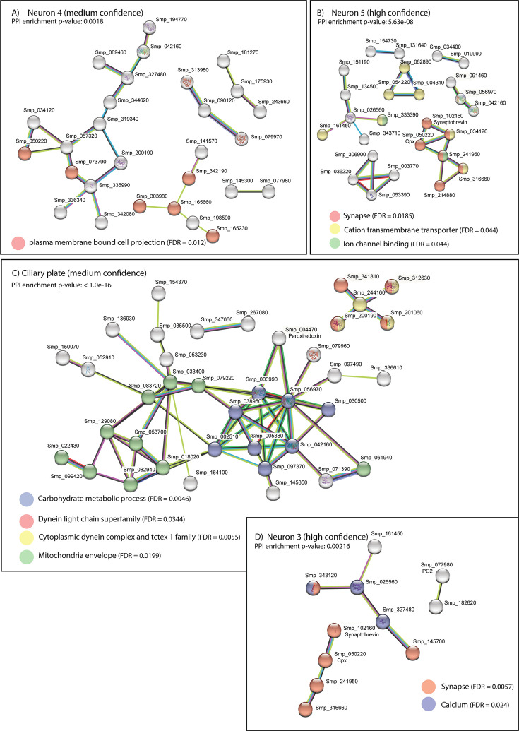
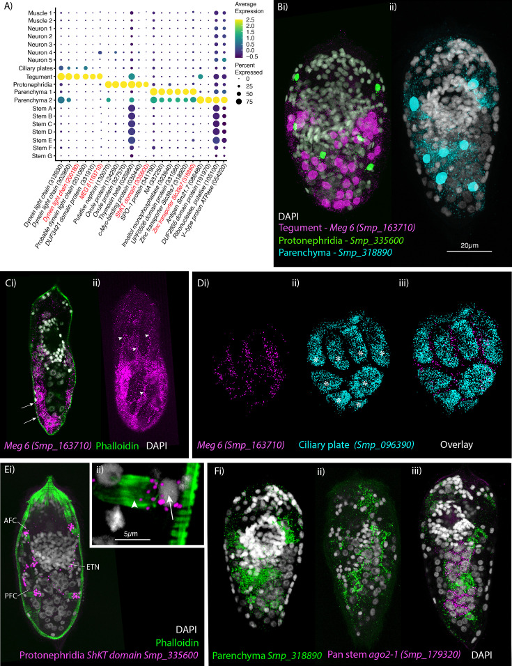
is a parasitic flatworm that causes the major neglected tropical disease schistosomiasis. The miracidium is the first larval stage of the life cycle. It swims and infects a freshwater snail, transforms into a mother sporocyst, where its stem cells generate daughter sporocysts that give rise to human-infective cercariae larvae. To understand the miracidium at cellular and molecular levels, we created a whole-body atlas of its ~365 cells. Single-cell RNA sequencing identified 19 transcriptionally distinct cell clusters. In situ hybridisation of tissue-specific genes revealed that 93% of the cells in the larva are somatic (57% neural, 19% muscle, 13% epidermal or tegument, 2% parenchyma, and 2% protonephridia) and 7% are stem. Whereas neurons represent the most diverse somatic cell types, trajectory analysis of the two main stem cell populations indicates that one of them is the origin of the tegument lineage and the other likely contains pluripotent cells. Furthermore, unlike the somatic cells, each of these stem populations shows sex-biased transcriptional signatures suggesting a cell-type-specific gene dosage compensation for sex chromosome-linked loci. The miracidium represents a simple developmental stage with which to gain a fundamental understanding of the molecular biology and spatial architecture of schistosome cells.

摘要

是一种寄生扁形动物,它会导致主要的被忽视热带病——血吸虫病。毛蚴是生命周期的第一个幼虫阶段。它在淡水中游泳并感染淡水螺,然后转化为母孢囊,其干细胞在母孢囊中生成可感染人类的尾蚴幼虫。为了在细胞和分子水平上了解毛蚴,我们创建了其约 365 个细胞的全身图谱。单细胞 RNA 测序鉴定出 19 个转录上不同的细胞簇。组织特异性基因的原位杂交显示,幼虫中的 93%的细胞是体细胞(57%是神经细胞,19%是肌肉细胞,13%是表皮或表皮细胞,2%是实质细胞,2%是原肾细胞),7%是干细胞。虽然神经元代表了最具多样性的体细胞类型,但对两个主要干细胞群的轨迹分析表明,其中一个是表皮谱系的起源,另一个可能含有多能细胞。此外,与体细胞不同,这些干细胞群体中的每一个都表现出性别偏倚的转录特征,这表明性染色体连锁基因座存在细胞类型特异性的基因剂量补偿。毛蚴代表了一个简单的发育阶段,可以帮助我们深入了解血吸虫细胞的分子生物学和空间结构。

https://cdn.ncbi.nlm.nih.gov/pmc/blobs/81b8/11349301/098d573b68df/elife-95628-fig5-figsupp2.jpg
https://cdn.ncbi.nlm.nih.gov/pmc/blobs/81b8/11349301/17491381c981/elife-95628-fig1.jpg
https://cdn.ncbi.nlm.nih.gov/pmc/blobs/81b8/11349301/6d358e63f7cb/elife-95628-fig1-figsupp1.jpg
https://cdn.ncbi.nlm.nih.gov/pmc/blobs/81b8/11349301/a1e0121cb20b/elife-95628-fig2.jpg
https://cdn.ncbi.nlm.nih.gov/pmc/blobs/81b8/11349301/f0d533ff2a97/elife-95628-fig3.jpg
https://cdn.ncbi.nlm.nih.gov/pmc/blobs/81b8/11349301/e31263c9fbda/elife-95628-fig3-figsupp1.jpg
https://cdn.ncbi.nlm.nih.gov/pmc/blobs/81b8/11349301/d44899454dec/elife-95628-fig3-figsupp2.jpg
https://cdn.ncbi.nlm.nih.gov/pmc/blobs/81b8/11349301/4b9d5f83897a/elife-95628-fig3-figsupp3.jpg
https://cdn.ncbi.nlm.nih.gov/pmc/blobs/81b8/11349301/24d5eafa3b4a/elife-95628-fig3-figsupp4.jpg
https://cdn.ncbi.nlm.nih.gov/pmc/blobs/81b8/11349301/20e689494c00/elife-95628-fig3-figsupp5.jpg
https://cdn.ncbi.nlm.nih.gov/pmc/blobs/81b8/11349301/c27126a49257/elife-95628-fig3-figsupp6.jpg
https://cdn.ncbi.nlm.nih.gov/pmc/blobs/81b8/11349301/1b1273293167/elife-95628-fig4.jpg
https://cdn.ncbi.nlm.nih.gov/pmc/blobs/81b8/11349301/d305ee7ae5f9/elife-95628-fig4-figsupp1.jpg
https://cdn.ncbi.nlm.nih.gov/pmc/blobs/81b8/11349301/23dcab602b2e/elife-95628-fig4-figsupp2.jpg
https://cdn.ncbi.nlm.nih.gov/pmc/blobs/81b8/11349301/ea254c2f88b7/elife-95628-fig4-figsupp3.jpg
https://cdn.ncbi.nlm.nih.gov/pmc/blobs/81b8/11349301/f6f23b655d5d/elife-95628-fig5-figsupp1.jpg
https://cdn.ncbi.nlm.nih.gov/pmc/blobs/81b8/11349301/098d573b68df/elife-95628-fig5-figsupp2.jpg

相似文献

1
A single-cell atlas of the miracidium larva of reveals cell types, developmental pathways, and tissue architecture.揭示尾蚴幼虫细胞类型、发育途径和组织架构的单细胞图谱。
Elife. 2024 Aug 27;13:RP95628. doi: 10.7554/eLife.95628.
2
Time series analysis of tegument ultrastructure of in vitro transformed miracidium to mother sporocyst of the human parasite Schistosoma mansoni.人体寄生虫曼氏血吸虫体外转化毛蚴至母胞蚴过程中皮层超微结构的时间序列分析。
Acta Trop. 2023 Apr;240:106840. doi: 10.1016/j.actatropica.2023.106840. Epub 2023 Jan 18.
3
Protein kinase C signalling during miracidium to mother sporocyst development in the helminth parasite, Schistosoma mansoni.蛋白激酶 C 在曼氏血吸虫从毛蚴向母胞蚴发育过程中的信号转导作用。
Int J Parasitol. 2009 Sep;39(11):1223-33. doi: 10.1016/j.ijpara.2009.04.002. Epub 2009 Apr 24.
4
Schistosoma mansoni: DNA microarray gene expression profiling during the miracidium-to-mother sporocyst transformation.曼氏血吸虫:从毛蚴到母胞蚴转变过程中的DNA微阵列基因表达谱分析
Mol Biochem Parasitol. 2006 May;147(1):39-47. doi: 10.1016/j.molbiopara.2006.01.006. Epub 2006 Feb 2.
5
Glycotope analysis in miracidia and primary sporocysts of Schistosoma mansoni: differential expression during the miracidium-to-sporocyst transformation.曼氏血吸虫毛蚴和初代孢囊的糖基表位分析:毛蚴向孢囊转化过程中的差异表达。
Int J Parasitol. 2009 Oct;39(12):1331-44. doi: 10.1016/j.ijpara.2009.06.002. Epub 2009 Jun 21.
6
Proteomic analysis of Schistosoma mansoni proteins released during in vitro miracidium-to-sporocyst transformation.曼氏血吸虫在体外从毛蚴转化为孢蚴过程中释放的蛋白质的蛋白质组学分析。
Mol Biochem Parasitol. 2009 Mar;164(1):32-44. doi: 10.1016/j.molbiopara.2008.11.005. Epub 2008 Nov 27.
7
Schistosoma mansoni: host cell adhesion to the different stages of the parasite, in vivo.曼氏血吸虫:体内宿主细胞与寄生虫不同阶段的黏附
Rev Inst Med Trop Sao Paulo. 1992 May-Jun;34(3):205-9. doi: 10.1590/s0036-46651992000300004.
8
Single-cell transcriptomics of the human parasite Schistosoma mansoni first intra-molluscan stage reveals tentative tegumental and stem-cell regulators.人类寄生虫曼氏血吸虫首个软体动物内阶段的单细胞转录组学揭示了初步的表皮和干细胞调节因子。
Sci Rep. 2024 Mar 12;14(1):5974. doi: 10.1038/s41598-024-55790-3.
9
Single-cell atlas of the first intra-mammalian developmental stage of the human parasite Schistosoma mansoni.人类寄生虫曼氏血吸虫在哺乳动物体内第一个发育阶段的单细胞图谱。
Nat Commun. 2020 Dec 18;11(1):6411. doi: 10.1038/s41467-020-20092-5.
10
A role for p38 mitogen-activated protein kinase in early post-embryonic development of Schistosoma mansoni.p38丝裂原活化蛋白激酶在曼氏血吸虫胚后早期发育中的作用。
Mol Biochem Parasitol. 2011 Nov;180(1):51-5. doi: 10.1016/j.molbiopara.2011.07.002. Epub 2011 Jul 19.

引用本文的文献

1
Tissue-Specific Distribution of Eggs in The Definitive Host Drives Transcriptomic and Behavioral Differences in Miracidia.终末宿主体内虫卵的组织特异性分布驱动毛蚴的转录组和行为差异。
bioRxiv. 2025 Aug 19:2025.08.15.670345. doi: 10.1101/2025.08.15.670345.
2
Establishing single cell RNA transcriptomics: a brief guide.建立单细胞RNA转录组学:简要指南。
Front Zool. 2025 Sep 2;22(1):25. doi: 10.1186/s12983-025-00579-x.
3
Quantitative ethology of schistosome miracidia characterizes a conserved snail peptide that inhibits penetration.

本文引用的文献

1
Single-cell transcriptomics of the human parasite Schistosoma mansoni first intra-molluscan stage reveals tentative tegumental and stem-cell regulators.人类寄生虫曼氏血吸虫首个软体动物内阶段的单细胞转录组学揭示了初步的表皮和干细胞调节因子。
Sci Rep. 2024 Mar 12;14(1):5974. doi: 10.1038/s41598-024-55790-3.
2
The stage- and sex-specific transcriptome of the human parasite Schistosoma mansoni.人体寄生虫曼氏血吸虫的阶段和性别特异性转录组。
Sci Data. 2023 Nov 7;10(1):775. doi: 10.1038/s41597-023-02674-2.
3
Fast and accurate protein structure search with Foldseek.
血吸虫毛蚴的定量行为学鉴定出一种保守的抑制穿透的蜗牛肽。
bioRxiv. 2025 Jul 3:2025.07.02.662618. doi: 10.1101/2025.07.02.662618.
4
Advances in Single-Cell Sequencing for Infectious Diseases: Progress and Perspectives.传染病单细胞测序的进展:现状与展望
Adv Sci (Weinh). 2025 Aug;12(32):e15678. doi: 10.1002/advs.202415678. Epub 2025 Jul 4.
5
How useful is Nanopore adaptive sampling for sequencing miracidia?纳米孔适应性采样在曼氏血吸虫毛蚴测序中作用有多大?
Wellcome Open Res. 2025 May 16;10:239. doi: 10.12688/wellcomeopenres.24094.1. eCollection 2025.
6
CRISPR/Cas genome editing, functional genomics, and diagnostics for parasitic helminths.用于寄生蠕虫的CRISPR/Cas基因组编辑、功能基因组学及诊断方法
Int J Parasitol. 2025 May 19. doi: 10.1016/j.ijpara.2025.05.001.
7
Eggs-posed: revision of Schistosoma mansoni venom allergen-like proteins unveils new genes and offers new insights into egg-host interactions.卵相关:曼氏血吸虫毒液过敏原样蛋白的修订揭示新基因并为卵与宿主相互作用提供新见解
BMC Genomics. 2025 Feb 24;26(1):189. doi: 10.1186/s12864-025-11369-4.
8
Regeneration in the absence of canonical neoblasts in an early branching flatworm.早期分支扁虫中缺乏典型新生细胞时的再生
Nat Commun. 2025 Jan 31;16(1):1232. doi: 10.1038/s41467-024-54716-x.
9
CRISPR-based functional genomics for schistosomes and related flatworms.基于 CRISPR 的血吸虫和相关扁形动物功能基因组学。
Trends Parasitol. 2024 Nov;40(11):1016-1028. doi: 10.1016/j.pt.2024.09.010. Epub 2024 Oct 18.
10
Regeneration in the absence of canonical neoblasts in an early branching flatworm.早期分支扁虫中缺乏典型新生细胞时的再生
bioRxiv. 2024 May 28:2024.05.24.595708. doi: 10.1101/2024.05.24.595708.
使用 Foldseek 进行快速准确的蛋白质结构搜索。
Nat Biotechnol. 2024 Feb;42(2):243-246. doi: 10.1038/s41587-023-01773-0. Epub 2023 May 8.
4
Time series analysis of tegument ultrastructure of in vitro transformed miracidium to mother sporocyst of the human parasite Schistosoma mansoni.人体寄生虫曼氏血吸虫体外转化毛蚴至母胞蚴过程中皮层超微结构的时间序列分析。
Acta Trop. 2023 Apr;240:106840. doi: 10.1016/j.actatropica.2023.106840. Epub 2023 Jan 18.
5
The Transcriptome of Developing Eggs Reveals Key Mediators in Pathogenesis and Life Cycle Propagation.发育中卵的转录组揭示了发病机制和生命周期传播中的关键介质。
Front Trop Dis. 2021 Aug 18;2:713123. doi: 10.3389/fitd.2021.713123.
6
Single-cell transcriptomics identifies conserved regulators of neuroglandular lineages.单细胞转录组学鉴定神经腺谱系保守调控因子。
Cell Rep. 2022 Sep 20;40(12):111370. doi: 10.1016/j.celrep.2022.111370.
7
Convergent evolution of a genotoxic stress response in a parasite-specific p53 homolog.寄生虫特异性 p53 同源物中遗传毒性应激反应的趋同进化。
Proc Natl Acad Sci U S A. 2022 Sep 13;119(37):e2205201119. doi: 10.1073/pnas.2205201119. Epub 2022 Sep 6.
8
Convergent evolution of venom gland transcriptomes across Metazoa.后生动物毒液腺转录组的趋同进化。
Proc Natl Acad Sci U S A. 2022 Jan 4;119(1). doi: 10.1073/pnas.2111392119.
9
Whole-body integration of gene expression and single-cell morphology.整体基因表达与单细胞形态的整合。
Cell. 2021 Sep 2;184(18):4819-4837.e22. doi: 10.1016/j.cell.2021.07.017. Epub 2021 Aug 10.
10
Molecular evidence for a single origin of ultrafiltration-based excretory organs.基于超滤作用的排泄器官单一起源的分子证据。
Curr Biol. 2021 Aug 23;31(16):3629-3638.e2. doi: 10.1016/j.cub.2021.05.057. Epub 2021 Jun 23.