• 文献检索
  • 文档翻译
  • 深度研究
  • 学术资讯
  • Suppr Zotero 插件Zotero 插件
  • 邀请有礼
  • 套餐&价格
  • 历史记录
应用&插件
Suppr Zotero 插件Zotero 插件浏览器插件Mac 客户端Windows 客户端微信小程序
定价
高级版会员购买积分包购买API积分包
服务
文献检索文档翻译深度研究API 文档MCP 服务
关于我们
关于 Suppr公司介绍联系我们用户协议隐私条款
关注我们

Suppr 超能文献

核心技术专利:CN118964589B侵权必究
粤ICP备2023148730 号-1Suppr @ 2026

文献检索

告别复杂PubMed语法,用中文像聊天一样搜索,搜遍4000万医学文献。AI智能推荐,让科研检索更轻松。

立即免费搜索

文件翻译

保留排版,准确专业,支持PDF/Word/PPT等文件格式,支持 12+语言互译。

免费翻译文档

深度研究

AI帮你快速写综述,25分钟生成高质量综述,智能提取关键信息,辅助科研写作。

立即免费体验

光学相干断层扫描在信号强度足够的情况下存在较高的伪影发生率。

High Prevalence of Artifacts in Optical Coherence Tomography With Adequate Signal Strength.

机构信息

Department of Ophthalmology, Casey Eye Institute, Oregon Health & Science University, Portland, OR, USA.

Department of Pharmacology and Neuroscience, North Texas Eye Research Institute, University of North Texas Health Science Center, Fort Worth, TX, USA.

出版信息

Transl Vis Sci Technol. 2024 Aug 1;13(8):43. doi: 10.1167/tvst.13.8.43.

DOI:10.1167/tvst.13.8.43
PMID:39196579
原文链接:https://pmc.ncbi.nlm.nih.gov/articles/PMC11364177/
Abstract

PURPOSE

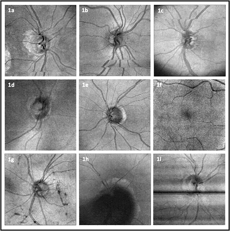
This study aims to investigate the prevalence of artifacts in optical coherence tomography (OCT) images with acceptable signal strength and evaluate the performance of supervised deep learning models in improving OCT image quality assessment.

METHODS

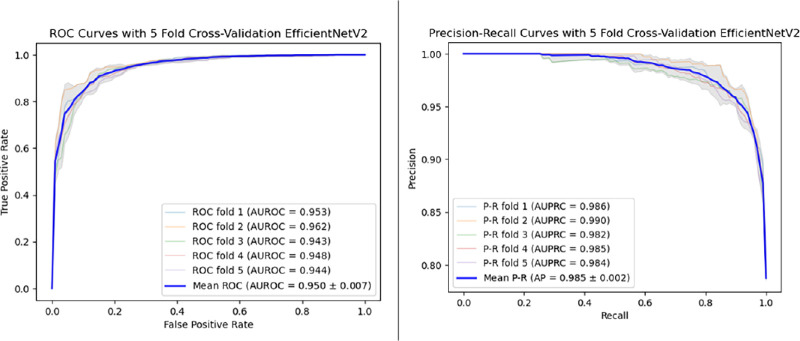
We conducted a retrospective study on 4555 OCT images from 546 patients, with each image having an acceptable signal strength (≥6). A comprehensive analysis of prevalent OCT artifacts was performed, and five pretrained convolutional neural network models were trained and tested to infer images based on quality.

RESULTS

Our results showed a high prevalence of artifacts in OCT images with acceptable signal strength. Approximately 21% of images were labeled as nonacceptable quality. The EfficientNetV2 model demonstrated superior performance in classifying OCT image quality, achieving an area under the receiver operating characteristic curve of 0.950 ± 0.007 and an area under the precision recall curve of 0.985 ± 0.002.

CONCLUSIONS

The findings highlight the limitations of relying solely on signal strength for OCT image quality assessment and the potential of deep learning models in accurately classifying image quality.

TRANSLATIONAL RELEVANCE

Application of the deep learning-based OCT image quality assessment models may improve the OCT image data quality for both clinical applications and research.

摘要

目的

本研究旨在调查具有可接受信号强度的光相干断层扫描(OCT)图像中的伪影发生率,并评估监督深度学习模型在改善 OCT 图像质量评估方面的性能。

方法

我们对 546 名患者的 4555 张 OCT 图像进行了回顾性研究,每张图像的信号强度均可接受(≥6)。我们对常见的 OCT 伪影进行了全面分析,并训练和测试了五个预先训练的卷积神经网络模型,以根据质量推断图像。

结果

我们的结果表明,具有可接受信号强度的 OCT 图像中存在很高的伪影发生率。大约 21%的图像被标记为质量不可接受。EfficientNetV2 模型在 OCT 图像质量分类方面表现出卓越的性能,其受试者工作特征曲线下面积为 0.950±0.007,精度召回曲线下面积为 0.985±0.002。

结论

这些发现强调了仅依赖信号强度评估 OCT 图像质量的局限性,以及深度学习模型在准确分类图像质量方面的潜力。

临床意义

基于深度学习的 OCT 图像质量评估模型的应用可能会提高 OCT 图像数据的质量,无论是在临床应用还是研究中。

https://cdn.ncbi.nlm.nih.gov/pmc/blobs/fd2c/11364177/28b0fcd99203/tvst-13-8-43-f002.jpg
https://cdn.ncbi.nlm.nih.gov/pmc/blobs/fd2c/11364177/7d40ee7d4bcb/tvst-13-8-43-f001.jpg
https://cdn.ncbi.nlm.nih.gov/pmc/blobs/fd2c/11364177/28b0fcd99203/tvst-13-8-43-f002.jpg
https://cdn.ncbi.nlm.nih.gov/pmc/blobs/fd2c/11364177/7d40ee7d4bcb/tvst-13-8-43-f001.jpg
https://cdn.ncbi.nlm.nih.gov/pmc/blobs/fd2c/11364177/28b0fcd99203/tvst-13-8-43-f002.jpg

相似文献

1
High Prevalence of Artifacts in Optical Coherence Tomography With Adequate Signal Strength.光学相干断层扫描在信号强度足够的情况下存在较高的伪影发生率。
Transl Vis Sci Technol. 2024 Aug 1;13(8):43. doi: 10.1167/tvst.13.8.43.
2
Self-Supervised Learning for Improved Optical Coherence Tomography Detection of Macular Telangiectasia Type 2.基于自监督学习的黄斑毛细血管扩张症 2 型光学相干断层扫描检测方法的研究
JAMA Ophthalmol. 2024 Mar 1;142(3):226-233. doi: 10.1001/jamaophthalmol.2023.6454.
3
SLO-Net: Enhancing Multiple Sclerosis Diagnosis Beyond Optical Coherence Tomography Using Infrared Reflectance Scanning Laser Ophthalmoscopy Images.SLO-Net:利用红外反射扫描激光检眼镜图像提升光学相干断层扫描技术之外的多发性硬化症诊断水平
Transl Vis Sci Technol. 2024 Jul 1;13(7):13. doi: 10.1167/tvst.13.7.13.
4
Development and Validation of a Convolutional Neural Network Model to Predict a Pathologic Fracture in the Proximal Femur Using Abdomen and Pelvis CT Images of Patients With Advanced Cancer.利用晚期癌症患者腹部和骨盆 CT 图像建立卷积神经网络模型预测股骨近端病理性骨折的研究
Clin Orthop Relat Res. 2023 Nov 1;481(11):2247-2256. doi: 10.1097/CORR.0000000000002771. Epub 2023 Aug 23.
5
Multi-Plexus Nonperfusion Area Segmentation in Widefield OCT Angiography Using a Deep Convolutional Neural Network.使用深度卷积神经网络对广角 OCT 血管造影中的多丛无灌注区进行分割。
Transl Vis Sci Technol. 2024 Jul 1;13(7):15. doi: 10.1167/tvst.13.7.15.
6
Optical coherence tomography (OCT) for detection of macular oedema in patients with diabetic retinopathy.光学相干断层扫描(OCT)用于检测糖尿病视网膜病变患者的黄斑水肿。
Cochrane Database Syst Rev. 2011 Jul 6(7):CD008081. doi: 10.1002/14651858.CD008081.pub2.
7
Novel Deep Learning Model for Glaucoma Detection Using Fusion of Fundus and Optical Coherence Tomography Images.基于眼底图像与光学相干断层扫描图像融合的青光眼检测新型深度学习模型
Sensors (Basel). 2025 Jul 11;25(14):4337. doi: 10.3390/s25144337.
8
Artificial intelligence for diagnosing exudative age-related macular degeneration.人工智能在渗出性年龄相关性黄斑变性诊断中的应用。
Cochrane Database Syst Rev. 2024 Oct 17;10(10):CD015522. doi: 10.1002/14651858.CD015522.pub2.
9
Optical coherence tomography (OCT) for detection of macular oedema in patients with diabetic retinopathy.光学相干断层扫描(OCT)用于检测糖尿病视网膜病变患者的黄斑水肿。
Cochrane Database Syst Rev. 2015 Jan 7;1(1):CD008081. doi: 10.1002/14651858.CD008081.pub3.
10
Deep learning generalization study on optical coherence tomography image denoising.光学相干断层扫描图像去噪的深度学习泛化研究
Phys Med Biol. 2025 Jun 25. doi: 10.1088/1361-6560/ade840.

引用本文的文献

1
Deep Learning and The Retina: A New Frontier in Multiple Sclerosis Diagnosis.深度学习与视网膜:多发性硬化症诊断的新前沿
Curr Health Sci J. 2025 Jan-Mar;51(1):26-36. doi: 10.12865/CHSJ.51.01.03. Epub 2025 Mar 31.
2
Artificial Intelligence for Optical Coherence Tomography in Glaucoma.用于青光眼光学相干断层扫描的人工智能
Transl Vis Sci Technol. 2025 Jan 2;14(1):27. doi: 10.1167/tvst.14.1.27.

本文引用的文献

1
Efficient and accurate compound scaling for convolutional neural networks.高效准确的卷积神经网络化合物缩放。
Neural Netw. 2023 Oct;167:787-797. doi: 10.1016/j.neunet.2023.08.053. Epub 2023 Sep 4.
2
The Prevalence of Optical Coherence Tomography Artifacts in High Myopia and its Influence on Glaucoma Diagnosis.高度近视患者光学相干断层扫描伪影的发生率及其对青光眼诊断的影响。
J Glaucoma. 2023 Sep 1;32(9):725-733. doi: 10.1097/IJG.0000000000002268. Epub 2023 Jul 19.
3
Deep learning in optical coherence tomography: Where are the gaps?
深度学习在光学相干断层扫描中的应用:存在哪些差距?
Clin Exp Ophthalmol. 2023 Nov;51(8):853-863. doi: 10.1111/ceo.14258. Epub 2023 May 28.
4
Deep Learning in Optical Coherence Tomography Angiography: Current Progress, Challenges, and Future Directions.光学相干断层扫描血管造影中的深度学习:当前进展、挑战及未来方向。
Diagnostics (Basel). 2023 Jan 16;13(2):326. doi: 10.3390/diagnostics13020326.
5
Deep learning for quality assessment of optical coherence tomography angiography images.深度学习在光学相干断层扫描血管造影图像质量评估中的应用。
Sci Rep. 2022 Aug 12;12(1):13775. doi: 10.1038/s41598-022-17709-8.
6
Deep Learning-Based Optical Coherence Tomography and Optical Coherence Tomography Angiography Image Analysis: An Updated Summary.基于深度学习的光学相干断层扫描和光学相干断层扫描血管造影图像分析:最新综述。
Asia Pac J Ophthalmol (Phila). 2021;10(3):253-260. doi: 10.1097/APO.0000000000000405.
7
Text Data Augmentation for Deep Learning.用于深度学习的文本数据增强
J Big Data. 2021;8(1):101. doi: 10.1186/s40537-021-00492-0. Epub 2021 Jul 19.
8
OCT Angiography Artifacts in Glaucoma.青光眼的 OCT 血管造影伪影。
Ophthalmology. 2021 Oct;128(10):1426-1437. doi: 10.1016/j.ophtha.2021.03.036. Epub 2021 Apr 2.
9
Artifacts in Macular Optical Coherence Tomography.黄斑光学相干断层扫描中的伪像
J Curr Ophthalmol. 2020 Apr 30;32(2):123-131. doi: 10.4103/JOCO.JOCO_83_20. eCollection 2020 Apr-Jun.
10
Prevalence and Severity of Artifacts in Optical Coherence Tomographic Angiograms.光学相干断层扫描血管造影术的病变发生率和严重程度。
JAMA Ophthalmol. 2020 Feb 1;138(2):119-126. doi: 10.1001/jamaophthalmol.2019.4971.