• 文献检索
  • 文档翻译
  • 深度研究
  • 学术资讯
  • Suppr Zotero 插件Zotero 插件
  • 邀请有礼
  • 套餐&价格
  • 历史记录
应用&插件
Suppr Zotero 插件Zotero 插件浏览器插件Mac 客户端Windows 客户端微信小程序
定价
高级版会员购买积分包购买API积分包
服务
文献检索文档翻译深度研究API 文档MCP 服务
关于我们
关于 Suppr公司介绍联系我们用户协议隐私条款
关注我们

Suppr 超能文献

核心技术专利:CN118964589B侵权必究
粤ICP备2023148730 号-1Suppr @ 2026

文献检索

告别复杂PubMed语法,用中文像聊天一样搜索,搜遍4000万医学文献。AI智能推荐,让科研检索更轻松。

立即免费搜索

文件翻译

保留排版,准确专业,支持PDF/Word/PPT等文件格式,支持 12+语言互译。

免费翻译文档

深度研究

AI帮你快速写综述,25分钟生成高质量综述,智能提取关键信息,辅助科研写作。

立即免费体验

深度学习在光学相干断层扫描血管造影图像质量评估中的应用。

Deep learning for quality assessment of optical coherence tomography angiography images.

机构信息

Department of Ophthalmology, Yale School of Medicine, New Haven, CT, 06510, USA.

Division of Oculoplastics and Reconstructive Surgery, Wilmer Eye Institute, Baltimore, MD, 21287, USA.

出版信息

Sci Rep. 2022 Aug 12;12(1):13775. doi: 10.1038/s41598-022-17709-8.

DOI:10.1038/s41598-022-17709-8
PMID:35962007
原文链接:https://pmc.ncbi.nlm.nih.gov/articles/PMC9374672/
Abstract

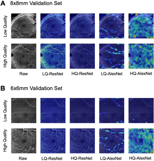
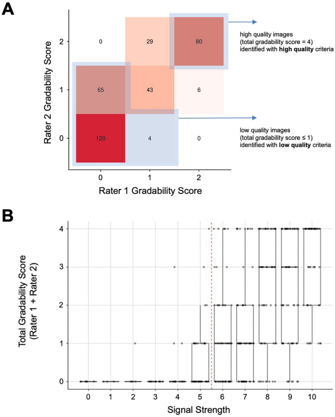
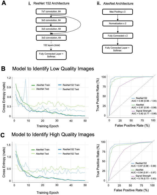
Optical coherence tomography angiography (OCTA) is an emerging non-invasive technique for imaging the retinal vasculature. While there are many promising clinical applications for OCTA, determination of image quality remains a challenge. We developed a deep learning-based system using a ResNet152 neural network classifier, pretrained using ImageNet, to classify images of the superficial capillary plexus in 347 scans from 134 patients. Images were also manually graded by two independent graders as a ground truth for the supervised learning models. Because requirements for image quality may vary depending on the clinical or research setting, two models were trained-one to identify high-quality images and one to identify low-quality images. Our neural network models demonstrated outstanding area under the curve (AUC) metrics for both low quality image identification (AUC = 0.99, 95%CI 0.98-1.00, [Formula: see text] = 0.90) and high quality image identification (AUC = 0.97, 95%CI 0.96-0.99, [Formula: see text] = 0.81), significantly outperforming machine-reported signal strength (AUC = 0.82, 95%CI 0.77-0.86, [Formula: see text]= 0.52 and AUC = 0.78, 95%CI 0.73-0.83, [Formula: see text] = 0.27 respectively). Our study demonstrates that techniques from machine learning may be used to develop flexible and robust methods for quality control of OCTA images.

摘要

光学相干断层扫描血管造影术(OCTA)是一种新兴的非侵入性技术,用于成像视网膜血管。虽然 OCTA 有许多有前途的临床应用,但图像质量的确定仍然是一个挑战。我们使用基于 ResNet152 神经网络分类器的深度学习系统,使用 ImageNet 进行预训练,对来自 134 名患者的 347 次扫描的浅层毛细血管丛图像进行分类。这些图像也由两名独立的评分者手动分级,作为监督学习模型的真实数据。由于图像质量的要求可能因临床或研究环境而异,因此我们训练了两个模型 - 一个用于识别高质量图像,另一个用于识别低质量图像。我们的神经网络模型在低质量图像识别(AUC = 0.99,95%CI 0.98-1.00,[Formula: see text] = 0.90)和高质量图像识别(AUC = 0.97,95%CI 0.96-0.99,[Formula: see text] = 0.81)方面均表现出出色的曲线下面积(AUC)指标,明显优于机器报告的信号强度(AUC = 0.82,95%CI 0.77-0.86,[Formula: see text]= 0.52 和 AUC = 0.78,95%CI 0.73-0.83,[Formula: see text] = 0.27)。我们的研究表明,机器学习技术可用于开发 OCTA 图像质量控制的灵活且强大的方法。

https://cdn.ncbi.nlm.nih.gov/pmc/blobs/861e/9374672/832a72591615/41598_2022_17709_Fig4_HTML.jpg
https://cdn.ncbi.nlm.nih.gov/pmc/blobs/861e/9374672/ae998d661b86/41598_2022_17709_Fig1_HTML.jpg
https://cdn.ncbi.nlm.nih.gov/pmc/blobs/861e/9374672/3d97c02e8ac6/41598_2022_17709_Fig2_HTML.jpg
https://cdn.ncbi.nlm.nih.gov/pmc/blobs/861e/9374672/5d3fbf980daf/41598_2022_17709_Fig3_HTML.jpg
https://cdn.ncbi.nlm.nih.gov/pmc/blobs/861e/9374672/832a72591615/41598_2022_17709_Fig4_HTML.jpg

相似文献

1
Deep learning for quality assessment of optical coherence tomography angiography images.深度学习在光学相干断层扫描血管造影图像质量评估中的应用。
Sci Rep. 2022 Aug 12;12(1):13775. doi: 10.1038/s41598-022-17709-8.
2
Automated OCT angiography image quality assessment using a deep learning algorithm.使用深度学习算法的自动光学相干断层扫描血管造影图像质量评估
Graefes Arch Clin Exp Ophthalmol. 2019 Aug;257(8):1641-1648. doi: 10.1007/s00417-019-04338-7. Epub 2019 May 22.
3
A MULTITASK DEEP-LEARNING SYSTEM FOR ASSESSMENT OF DIABETIC MACULAR ISCHEMIA ON OPTICAL COHERENCE TOMOGRAPHY ANGIOGRAPHY IMAGES.基于光学相干断层扫描血管造影图像的糖尿病性黄斑缺血评估的多任务深度学习系统。
Retina. 2022 Jan 1;42(1):184-194. doi: 10.1097/IAE.0000000000003287.
4
Ensemble Deep Learning for Diabetic Retinopathy Detection Using Optical Coherence Tomography Angiography.基于光学相干断层扫描血管造影的糖尿病视网膜病变检测的集成深度学习。
Transl Vis Sci Technol. 2020 Apr 13;9(2):20. doi: 10.1167/tvst.9.2.20. eCollection 2020 Apr.
5
Automated Diagnosis of Optical Coherence Tomography Angiography (OCTA) Based on Machine Learning Techniques.基于机器学习技术的光学相干断层扫描血管造影(OCTA)自动诊断。
Sensors (Basel). 2022 Mar 18;22(6):2342. doi: 10.3390/s22062342.
6
Machine learning in optical coherence tomography angiography.光学相干断层扫描血管造影中的机器学习。
Exp Biol Med (Maywood). 2021 Oct;246(20):2170-2183. doi: 10.1177/15353702211026581. Epub 2021 Jul 19.
7
A novel deep learning conditional generative adversarial network for producing angiography images from retinal fundus photographs.一种新颖的深度学习条件生成对抗网络,用于从眼底照片生成血管造影图像。
Sci Rep. 2020 Dec 9;10(1):21580. doi: 10.1038/s41598-020-78696-2.
8
A Deep Learning Algorithm for Classifying Diabetic Retinopathy Using Optical Coherence Tomography Angiography.基于光学相干断层扫描血管造影的糖尿病视网膜病变深度学习算法分类。
Transl Vis Sci Technol. 2022 Feb 1;11(2):39. doi: 10.1167/tvst.11.2.39.
9
Automated detection of a nonperfusion area caused by retinal vein occlusion in optical coherence tomography angiography images using deep learning.利用深度学习技术在光学相干断层扫描血管造影图像中自动检测视网膜静脉阻塞引起的无灌注区。
PLoS One. 2019 Nov 7;14(11):e0223965. doi: 10.1371/journal.pone.0223965. eCollection 2019.
10
Diagnosing Diabetic Retinopathy in OCTA Images Based on Multilevel Information Fusion Using a Deep Learning Framework.基于深度学习框架的多级信息融合在 OCTA 图像中诊断糖尿病视网膜病变。
Comput Math Methods Med. 2022 Aug 4;2022:4316507. doi: 10.1155/2022/4316507. eCollection 2022.

引用本文的文献

1
High Prevalence of Artifacts in Optical Coherence Tomography With Adequate Signal Strength.光学相干断层扫描在信号强度足够的情况下存在较高的伪影发生率。
Transl Vis Sci Technol. 2024 Aug 1;13(8):43. doi: 10.1167/tvst.13.8.43.
2
Convolutional Neural Network-Based Automated Quality Assessment of OCT and OCT Angiography Image Maps in Individuals With Neurodegenerative Disease.基于卷积神经网络的神经退行性疾病患者 OCT 和 OCT 血管造影图像地图的自动质量评估。
Transl Vis Sci Technol. 2023 Jun 1;12(6):30. doi: 10.1167/tvst.12.6.30.
3
Optical Coherence Tomography Angiography of the Intestine: How to Prevent Motion Artifacts in Open and Laparoscopic Surgery?

本文引用的文献

1
Text Data Augmentation for Deep Learning.用于深度学习的文本数据增强
J Big Data. 2021;8(1):101. doi: 10.1186/s40537-021-00492-0. Epub 2021 Jul 19.
2
Optical Coherence Tomography Angiography in Diabetes and Diabetic Retinopathy.糖尿病及糖尿病视网膜病变中的光学相干断层扫描血管造影
J Clin Med. 2020 Jun 3;9(6):1723. doi: 10.3390/jcm9061723.
3
Use of OCTA Capillary Perfusion Density Measurements to Detect and Grade Macular Ischemia.利用 OCTA 毛细血管灌注密度测量来检测和分级黄斑缺血。
肠道光学相干断层扫描血管造影:如何在开放手术和腹腔镜手术中预防运动伪影?
Life (Basel). 2023 Mar 6;13(3):705. doi: 10.3390/life13030705.
4
Deep Learning in Optical Coherence Tomography Angiography: Current Progress, Challenges, and Future Directions.光学相干断层扫描血管造影中的深度学习:当前进展、挑战及未来方向。
Diagnostics (Basel). 2023 Jan 16;13(2):326. doi: 10.3390/diagnostics13020326.
5
Using Artificial Intelligence to Analyse the Retinal Vascular Network: The Future of Cardiovascular Risk Assessment Based on Oculomics? A Narrative Review.利用人工智能分析视网膜血管网络:基于眼科学的心血管风险评估的未来?一篇综述。
Ophthalmol Ther. 2023 Apr;12(2):657-674. doi: 10.1007/s40123-022-00641-5. Epub 2022 Dec 23.
Ophthalmic Surg Lasers Imaging Retina. 2020 Apr 1;51(4):S30-S36. doi: 10.3928/23258160-20200401-04.
4
Imaging Artifacts and Segmentation Errors With Wide-Field Swept-Source Optical Coherence Tomography Angiography in Diabetic Retinopathy.糖尿病视网膜病变中广角扫频源光学相干断层扫描血管造影的影像伪影与分割误差
Transl Vis Sci Technol. 2019 Nov 15;8(6):18. doi: 10.1167/tvst.8.6.18. eCollection 2019 Nov.
5
Signal Strength Reduction Effects in OCT Angiography.光学相干断层扫描血管造影中的信号强度降低效应
Ophthalmol Retina. 2019 Oct;3(10):835-842. doi: 10.1016/j.oret.2019.04.029. Epub 2019 May 8.
6
Automated OCT angiography image quality assessment using a deep learning algorithm.使用深度学习算法的自动光学相干断层扫描血管造影图像质量评估
Graefes Arch Clin Exp Ophthalmol. 2019 Aug;257(8):1641-1648. doi: 10.1007/s00417-019-04338-7. Epub 2019 May 22.
7
Transfer Learning with Deep Convolutional Neural Networks for Classifying Cellular Morphological Changes.基于深度卷积神经网络的迁移学习在细胞形态变化分类中的应用
SLAS Discov. 2019 Apr;24(4):466-475. doi: 10.1177/2472555218818756. Epub 2019 Jan 14.
8
The Importance of Signal Strength in Quantitative Assessment of Retinal Vessel Density Using Optical Coherence Tomography Angiography.光学相干断层扫描血管造影定量评估视网膜血管密度中信号强度的重要性。
Sci Rep. 2018 Aug 27;8(1):12897. doi: 10.1038/s41598-018-31321-9.
9
Prevalences of segmentation errors and motion artifacts in OCT-angiography differ among retinal diseases.光学相干断层扫描血管造影(OCT-A)中分割误差和运动伪影的患病率在不同视网膜疾病中存在差异。
Graefes Arch Clin Exp Ophthalmol. 2018 Oct;256(10):1807-1816. doi: 10.1007/s00417-018-4053-2. Epub 2018 Jul 7.
10
Identification of imaging features that determine quality and repeatability of retinal capillary plexus density measurements in OCT angiography.确定影响 OCT 血管造影中视网膜毛细血管丛密度测量质量和可重复性的影像学特征。
Br J Ophthalmol. 2018 Apr;102(4):509-514. doi: 10.1136/bjophthalmol-2017-310700. Epub 2017 Aug 16.