• 文献检索
  • 文档翻译
  • 深度研究
  • 学术资讯
  • Suppr Zotero 插件Zotero 插件
  • 邀请有礼
  • 套餐&价格
  • 历史记录
应用&插件
Suppr Zotero 插件Zotero 插件浏览器插件Mac 客户端Windows 客户端微信小程序
定价
高级版会员购买积分包购买API积分包
服务
文献检索文档翻译深度研究API 文档MCP 服务
关于我们
关于 Suppr公司介绍联系我们用户协议隐私条款
关注我们

Suppr 超能文献

核心技术专利:CN118964589B侵权必究
粤ICP备2023148730 号-1Suppr @ 2026

文献检索

告别复杂PubMed语法,用中文像聊天一样搜索,搜遍4000万医学文献。AI智能推荐,让科研检索更轻松。

立即免费搜索

文件翻译

保留排版,准确专业,支持PDF/Word/PPT等文件格式,支持 12+语言互译。

免费翻译文档

深度研究

AI帮你快速写综述,25分钟生成高质量综述,智能提取关键信息,辅助科研写作。

立即免费体验

相似文献

1
Effect of Spectral Filtering and Segmental X-ray Tube Current Switch-Off on Interventionalist's Scatter Exposure during CT Fluoroscopy.光谱滤波和分段X射线管电流关闭对CT透视期间介入医生散射剂量的影响。
Bioengineering (Basel). 2024 Aug 16;11(8):838. doi: 10.3390/bioengineering11080838.
2
CT Fluoroscopy for Image-Guided Procedures: Physician Radiation Dose During Full-Rotation and Partial-Angle CT Scanning.CT 透视在影像引导操作中的应用:全旋转和部分角度 CT 扫描时医师的辐射剂量。
J Vasc Interv Radiol. 2021 Mar;32(3):439-446. doi: 10.1016/j.jvir.2020.10.033. Epub 2021 Jan 4.
3
The importance of spectral separation: an assessment of dual-energy spectral separation for quantitative ability and dose efficiency.光谱分离的重要性:对双能光谱分离的定量能力和剂量效率的评估。
Invest Radiol. 2015 Feb;50(2):114-8. doi: 10.1097/RLI.0000000000000109.
4
The effect of tube voltage combination on image artefact and radiation dose in dual-source dual-energy CT: comparison between conventional 80/140 kV and 80/150 kV plus tin filter for gout protocol.管电压组合对双源双能 CT 图像伪影和辐射剂量的影响:常规 80/140kV 与 80/150kV 加锡滤器在痛风方案中的比较。
Eur Radiol. 2019 Mar;29(3):1248-1257. doi: 10.1007/s00330-018-5622-9. Epub 2018 Jul 9.
5
Computed tomography arthrography of the shoulder with tin filter-based spectral shaping at 100 kV and 140 kV.在100 kV和140 kV下使用基于锡滤过器的光谱整形技术对肩部进行计算机断层扫描关节造影。
Acta Radiol. 2021 Oct;62(10):1349-1357. doi: 10.1177/0284185120965551. Epub 2020 Oct 18.
6
Ultra-low dose CT scanning for PET/CT.用于 PET/CT 的超低剂量 CT 扫描。
Med Phys. 2024 Jan;51(1):139-155. doi: 10.1002/mp.16862. Epub 2023 Dec 4.
7
Radiation exposure of the radiologist's eye lens during CT-guided interventions.CT引导介入操作过程中放射科医生晶状体的辐射暴露。
Acta Radiol. 2014 Feb;55(1):86-90. doi: 10.1177/0284185113493222. Epub 2013 Jul 24.
8
Technical Note: Radiation dose reduction from computed tomography localizer radiographs using a tin spectral shaping filter.技术说明:使用锡谱整形滤波器降低计算机断层定位摄影的辐射剂量。
Med Phys. 2019 Feb;46(2):544-549. doi: 10.1002/mp.13353. Epub 2019 Jan 15.
9
Radiation dose and image conspicuity comparison between conventional 120 kVp and 150 kVp with spectral beam shaping for temporal bone CT.常规 120 kVp 和 150 kVp 与带能谱成型技术 tempora 骨 CT 辐射剂量和图像显示的比较。
Eur J Radiol. 2018 May;102:68-73. doi: 10.1016/j.ejrad.2018.03.004. Epub 2018 Mar 7.
10
Dose Reduction for Sinus and Temporal Bone Imaging Using Photon-Counting Detector CT With an Additional Tin Filter.使用带附加锡滤过器的光子计数探测器 CT 进行鼻窦和颞骨成像的剂量降低。
Invest Radiol. 2020 Feb;55(2):91-100. doi: 10.1097/RLI.0000000000000614.

本文引用的文献

1
Radiation protection in CT-guided interventions: does real-time dose visualisation lead to a reduction in radiation dose to participating radiologists? A single-centre evaluation.CT 引导介入中的辐射防护:实时剂量可视化是否会降低参与放射科医生的辐射剂量?一项单中心评估。
Clin Radiol. 2024 Jun;79(6):e785-e790. doi: 10.1016/j.crad.2024.01.028. Epub 2024 Feb 15.
2
Radioprotective Effects of a Semicircular X-ray Shielding Device for Operators During CT Fluoroscopy-Guided Interventional Procedures: Experimental and Clinical Studies.用于 CT 透视引导介入手术中操作人员的半圆形 X 射线屏蔽装置的放射防护效果:实验与临床研究。
Cardiovasc Intervent Radiol. 2023 Jun;46(6):770-776. doi: 10.1007/s00270-023-03456-4. Epub 2023 May 15.
3
Comparative analysis of patient scatter to phantom scatter for computed tomography systems.计算机断层扫描系统中患者散射与体模散射的对比分析。
Phys Eng Sci Med. 2022 Sep;45(3):883-888. doi: 10.1007/s13246-022-01158-8. Epub 2022 Aug 3.
4
Radioablation of Upper Abdominal Malignancies by CT-Guided, Interstitial HDR Brachytherapy: A Multivariate Analysis of Catheter Placement Assisted by Ultrasound Imaging.
Rofo. 2022 Jan;194(1):62-69. doi: 10.1055/a-1545-4983. Epub 2021 Oct 14.
5
CIRSE Clinical Practice Manual.CIRSE 临床实践手册。
Cardiovasc Intervent Radiol. 2021 Sep;44(9):1323-1353. doi: 10.1007/s00270-021-02904-3. Epub 2021 Jul 6.
6
Cutting Staff Radiation Exposure and Improving Freedom of Motion during CT Interventions: Comparison of a Novel Workflow Utilizing a Radiation Protection Cabin versus Two Conventional Workflows.降低CT介入操作期间工作人员的辐射暴露并改善活动自由度:使用辐射防护舱的新型工作流程与两种传统工作流程的比较
Diagnostics (Basel). 2021 Jun 16;11(6):1099. doi: 10.3390/diagnostics11061099.
7
Occupational Radiation Dose to Eye Lenses in CT-Guided Interventions Using MDCT-Fluoroscopy.多层螺旋CT透视引导下介入操作中眼晶状体的职业辐射剂量
Diagnostics (Basel). 2021 Apr 2;11(4):646. doi: 10.3390/diagnostics11040646.
8
CT Fluoroscopy for Image-Guided Procedures: Physician Radiation Dose During Full-Rotation and Partial-Angle CT Scanning.CT 透视在影像引导操作中的应用:全旋转和部分角度 CT 扫描时医师的辐射剂量。
J Vasc Interv Radiol. 2021 Mar;32(3):439-446. doi: 10.1016/j.jvir.2020.10.033. Epub 2021 Jan 4.
9
Radiation dose optimization in diagnostic and interventional radiology: Current issues and future perspectives.诊断和介入放射学中的辐射剂量优化:当前问题和未来展望。
Phys Med. 2020 Nov;79:16-21. doi: 10.1016/j.ejmp.2020.09.015. Epub 2020 Oct 6.
10
Impact of interventionalist's experience and gender on radiation dose and procedural time in CT-guided interventions-a retrospective analysis of 4380 cases over 10 years.介入医生经验和性别对 CT 引导介入治疗中辐射剂量和手术时间的影响-一项回顾性分析,涉及 10 年中的 4380 例病例。
Eur Radiol. 2021 Feb;31(2):569-579. doi: 10.1007/s00330-020-07185-x. Epub 2020 Aug 26.

光谱滤波和分段X射线管电流关闭对CT透视期间介入医生散射剂量的影响。

Effect of Spectral Filtering and Segmental X-ray Tube Current Switch-Off on Interventionalist's Scatter Exposure during CT Fluoroscopy.

作者信息

Grosser Oliver S, Volk Martin, Georgiades Marilena, Punzet Daniel, Alsawalhi Bahaa, Kupitz Dennis, Omari Jazan, Wissel Heiko, Kreissl Michael C, Rose Georg, Pech Maciej

机构信息

Department of Radiology and Nuclear Medicine, University Hospital Magdeburg and Medical Faculty of Otto-von-Guericke University, 39120 Magdeburg, Germany.

Research Campus STIMULATE, Otto-von-Guericke University, 39106 Magdeburg, Germany.

出版信息

Bioengineering (Basel). 2024 Aug 16;11(8):838. doi: 10.3390/bioengineering11080838.

DOI:10.3390/bioengineering11080838
PMID:39199796
原文链接:https://pmc.ncbi.nlm.nih.gov/articles/PMC11351108/
Abstract

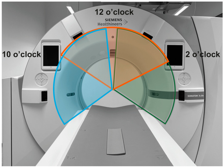
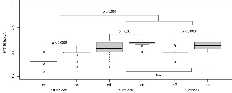
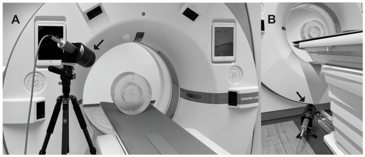
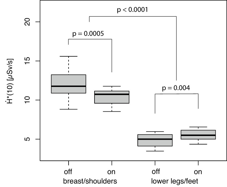
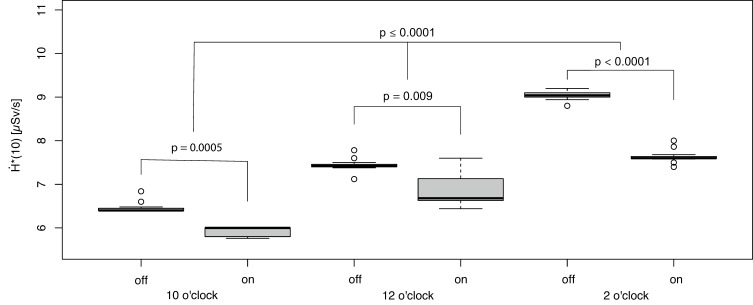
Dose optimization in computed tomography (CT) is crucial, especially in CT fluoroscopy (fluoro-CT) used for real-time navigation, affecting both patient and operator safety. This study evaluated the impact of spectral X-ray filtering using a tin filter (Sn filter), and a method called partial-angle computed tomography (PACT), which involves segmentally switching off the X-ray tube current at the ambient dose rate H˙(10) at the interventional radiologist's (IR) position. Measurements were taken at two body regions (upper body: head/neck; lower body: lower legs/feet) using a 120 kV X-ray tube voltage, 3 × 5.0 mm CT collimation, 0.5 s rotation speed, and X-ray tube currents of 43 Eff.mAs (without Sn filter) and 165 Eff.mAs (with Sn filter). The study found significant dose reductions in both body regions when using the Sn filter and PACT together. For instance, in the upper body region, the combination protocol reduced H˙(10) from 11.8 µSv/s to 6.1 µSv/s ( < 0.0001) compared to the protocol without using these features. Around 8% of the reduction (about 0.5 µSv/s) is attributed to the Sn filter ( = 0.0005). This approach demonstrates that using the Sn filter along with PACT effectively minimizes radiation exposure for the IR, particularly protecting areas like the head/neck, which can only be insufficiently covered by (standard) radiation protection material.

摘要

计算机断层扫描(CT)中的剂量优化至关重要,尤其是在用于实时导航的CT透视(荧光CT)中,这会影响患者和操作人员的安全。本研究评估了使用锡滤过器(Sn滤过器)进行光谱X射线滤过以及一种称为部分角度计算机断层扫描(PACT)的方法的影响,该方法涉及在介入放射科医生(IR)位置的环境剂量率H˙(10)下分段关闭X射线管电流。使用120 kV X射线管电压、3×5.0 mm CT准直、0.5 s旋转速度以及43等效毫安秒(无Sn滤过器)和165等效毫安秒(有Sn滤过器)的X射线管电流,在两个身体部位(上半身:头部/颈部;下半身:小腿/足部)进行测量。研究发现,同时使用Sn滤过器和PACT时,两个身体部位的剂量均显著降低。例如,在上半身区域,与未使用这些功能的方案相比,联合方案将H˙(10)从11.8 µSv/s降低至6.1 µSv/s(<0.0001)。约8%的降低(约0.5 µSv/s)归因于Sn滤过器(=0.0005)。这种方法表明,将Sn滤过器与PACT一起使用可有效降低IR的辐射暴露,特别是对头/颈部等(标准)辐射防护材料覆盖不足的区域起到保护作用。

https://cdn.ncbi.nlm.nih.gov/pmc/blobs/72ed/11351108/3e69a7fac453/bioengineering-11-00838-g005.jpg
https://cdn.ncbi.nlm.nih.gov/pmc/blobs/72ed/11351108/d871d93eaa2b/bioengineering-11-00838-g001.jpg
https://cdn.ncbi.nlm.nih.gov/pmc/blobs/72ed/11351108/9b357c2a7343/bioengineering-11-00838-g002.jpg
https://cdn.ncbi.nlm.nih.gov/pmc/blobs/72ed/11351108/c18b5147ac79/bioengineering-11-00838-g003.jpg
https://cdn.ncbi.nlm.nih.gov/pmc/blobs/72ed/11351108/a3fb4f75ef4e/bioengineering-11-00838-g004.jpg
https://cdn.ncbi.nlm.nih.gov/pmc/blobs/72ed/11351108/3e69a7fac453/bioengineering-11-00838-g005.jpg