Shen Xinyi, Zhu Zhengyang, Li Xin, Zhang Wen, Zhang Xin, Zhang Bing
Department of Radiology, Affiliated Hospital of Medical School, Nanjing Drum Tower Hospital, Nanjing University, Nanjing, China.
Institute of Medical Imaging and Artificial Intelligence, Nanjing University, Nanjing, China.
Front Neurosci. 2024 Aug 14;18:1442176. doi: 10.3389/fnins.2024.1442176. eCollection 2024.
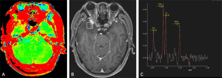
Cerebral syphilitic gumma is a rare intracranial infectious disorder. Without a clear history of syphilis and comprehensive serological examinations, it's challenging to diagnose it accurately prior to surgery through routine magnetic resonance imaging (MRI). Advanced neuroimaging techniques have been widely used in diagnosing brain tumors, yet there's limited report on their application in cerebral syphilitic gumma. This report presents a case of an elderly male patient with cerebral syphilitic gumma and analyzes its characteristics of advanced neuroimaging.
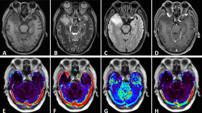
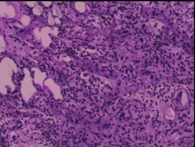
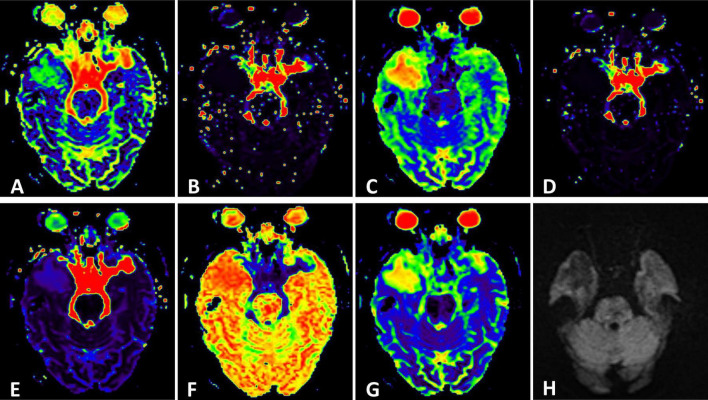
A 68-year-old male patient was admitted to our institution presenting with bilateral hearing loss complicated with continuing headaches without obvious cause. Laboratory tests indicated positive treponema pallidum. Conventional MRI showed nodules closely related to the adjacent meninges in bilateral temporal lobes. The patient underwent surgical resection of the nodule in the right temporal lobe due to the mass effect and the final pathological diagnosis revealed cerebral syphilitic gumma.
With the return of syphilis in recent years, accurate diagnosis of cerebral syphilitic gumma is a matter of great urgency. Advanced neuro-MRI can serve as a significant complement to conventional MRI examination.
脑梅毒瘤是一种罕见的颅内感染性疾病。在没有明确梅毒病史和全面血清学检查的情况下,术前通过常规磁共振成像(MRI)准确诊断具有挑战性。先进的神经影像学技术已广泛应用于脑肿瘤的诊断,但在脑梅毒瘤中的应用报道有限。本报告介绍了一例老年男性脑梅毒瘤患者,并分析了其先进神经影像学特征。
一名68岁男性患者因双侧听力丧失并伴有原因不明的持续性头痛入住我院。实验室检查显示梅毒螺旋体阳性。常规MRI显示双侧颞叶有与相邻脑膜密切相关的结节。由于占位效应,患者接受了右侧颞叶结节的手术切除,最终病理诊断为脑梅毒瘤。
近年来随着梅毒的再度出现,准确诊断脑梅毒瘤迫在眉睫。先进的神经MRI可作为常规MRI检查的重要补充。