• 文献检索
  • 文档翻译
  • 深度研究
  • 学术资讯
  • Suppr Zotero 插件Zotero 插件
  • 邀请有礼
  • 套餐&价格
  • 历史记录
应用&插件
Suppr Zotero 插件Zotero 插件浏览器插件Mac 客户端Windows 客户端微信小程序
定价
高级版会员购买积分包购买API积分包
服务
文献检索文档翻译深度研究API 文档MCP 服务
关于我们
关于 Suppr公司介绍联系我们用户协议隐私条款
关注我们

Suppr 超能文献

核心技术专利:CN118964589B侵权必究
粤ICP备2023148730 号-1Suppr @ 2026

文献检索

告别复杂PubMed语法,用中文像聊天一样搜索,搜遍4000万医学文献。AI智能推荐,让科研检索更轻松。

立即免费搜索

文件翻译

保留排版,准确专业,支持PDF/Word/PPT等文件格式,支持 12+语言互译。

免费翻译文档

深度研究

AI帮你快速写综述,25分钟生成高质量综述,智能提取关键信息,辅助科研写作。

立即免费体验

发育中的海马体:从童年到青少年期的微观结构演变

The developing hippocampus: Microstructural evolution through childhood and adolescence.

作者信息

Karat Bradley G, Genc Sila, Raven Erika P, Palombo Marco, Khan Ali R, Jones Derek K

机构信息

Robarts Research Institute, Western University, London, ON, Canada.

Centre for Functional and Metabolic Mapping, Western University, London, ON, Canada.

出版信息

bioRxiv. 2024 Aug 19:2024.08.19.608590. doi: 10.1101/2024.08.19.608590.

DOI:10.1101/2024.08.19.608590
PMID:39229062
原文链接:https://pmc.ncbi.nlm.nih.gov/articles/PMC11370384/
Abstract

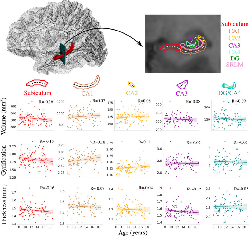
The hippocampus is a structure in the medial temporal lobe which serves multiple cognitive functions. While important, the development of the hippocampus in the formative period of childhood and adolescence has not been extensively investigated, with most contemporary research focusing on macrostructural measures of volume. Thus, there has been little research on the development of the micron-scale structures (i.e., microstructure) of the hippocampus, which engender its cognitive functions. The current study examined age-related changes of hippocampal microstructure using diffusion MRI data acquired with an ultra-strong gradient (300 mT/m) MRI scanner in a sample of children and adolescents (N=88; 8-19 years). Surface-based hippocampal modelling was combined with established microstructural approaches, such as Diffusion Tensor Imaging (DTI) and Neurite Orientation Dispersion Density Imaging (NODDI), and a more advanced gray matter diffusion model Soma And Neurite Density Imaging (SANDI). No significant changes in macrostructural measures (volume, gyrification, and thickness) were found between 8-19 years, while significant changes in microstructure measures related to neurites (from NODDI and SANDI), soma (from SANDI), and mean diffusivity (from DTI) were found. In particular, there was a significant increase across age in neurite MR signal fraction and a significant decrease in extracellular MR signal fraction and mean diffusivity across the hippocampal subfields and long-axis. A significant negative correlation between age and MR apparent soma radius was found in the subiculum and CA1 throughout the anterior and body of the hippocampus. Further surface-based analyses uncovered variability in age-related microstructural changes between the subfields and long-axis, which may reflect ostensible developmental differences along these two axes. Finally, correlation of hippocampal surfaces representing age-related changes of microstructure with maps derived from histology allowed for postulation of the potential underlying microstructure that diffusion changes across age may be capturing. Overall, distinct neurite and soma developmental profiles in the human hippocampus during late childhood and adolescence are reported for the first time.

摘要

海马体是内侧颞叶中的一种结构,具有多种认知功能。虽然海马体很重要,但在儿童期和青少年期的形成阶段,其发育情况尚未得到广泛研究,当代大多数研究都集中在体积的宏观结构测量上。因此,关于海马体微观尺度结构(即微结构)的发育研究很少,而正是这些微结构产生了其认知功能。本研究使用超强梯度(300 mT/m)MRI扫描仪采集的扩散MRI数据,对儿童和青少年样本(N = 88;8 - 19岁)中海马体微结构的年龄相关变化进行了研究。基于表面的海马体建模与既定的微结构方法相结合,如扩散张量成像(DTI)和神经突方向分散密度成像(NODDI),以及一种更先进的灰质扩散模型——体细胞和神经突密度成像(SANDI)。在8 - 19岁之间,未发现宏观结构测量指标(体积、脑回形成和厚度)有显著变化,而在与神经突(来自NODDI和SANDI)、体细胞(来自SANDI)和平均扩散率(来自DTI)相关的微结构测量指标上发现了显著变化。特别是,神经突MR信号分数随年龄显著增加,细胞外MR信号分数和平均扩散率在海马体亚区和长轴上显著降低。在海马体前部和体部的整个下托和CA1区域,发现年龄与MR表观体细胞半径之间存在显著负相关。进一步基于表面的分析揭示了亚区和长轴之间与年龄相关的微结构变化的变异性,这可能反映了沿这两个轴表面上的发育差异。最后,将代表微结构年龄相关变化的海马体表与组织学图谱进行相关性分析,有助于推测扩散随年龄变化可能捕捉到的潜在微结构。总体而言,首次报告了人类海马体在儿童晚期和青少年期独特的神经突和体细胞发育特征。

https://cdn.ncbi.nlm.nih.gov/pmc/blobs/c8d9/11370384/07651718250e/nihpp-2024.08.19.608590v1-f0009.jpg
https://cdn.ncbi.nlm.nih.gov/pmc/blobs/c8d9/11370384/b1e81e8f8c4d/nihpp-2024.08.19.608590v1-f0001.jpg
https://cdn.ncbi.nlm.nih.gov/pmc/blobs/c8d9/11370384/a272ea806025/nihpp-2024.08.19.608590v1-f0002.jpg
https://cdn.ncbi.nlm.nih.gov/pmc/blobs/c8d9/11370384/404c09ceeb18/nihpp-2024.08.19.608590v1-f0003.jpg
https://cdn.ncbi.nlm.nih.gov/pmc/blobs/c8d9/11370384/161b98fbaab5/nihpp-2024.08.19.608590v1-f0004.jpg
https://cdn.ncbi.nlm.nih.gov/pmc/blobs/c8d9/11370384/561ab7ad8533/nihpp-2024.08.19.608590v1-f0005.jpg
https://cdn.ncbi.nlm.nih.gov/pmc/blobs/c8d9/11370384/fccbb8532a3b/nihpp-2024.08.19.608590v1-f0006.jpg
https://cdn.ncbi.nlm.nih.gov/pmc/blobs/c8d9/11370384/126157f4c59e/nihpp-2024.08.19.608590v1-f0007.jpg
https://cdn.ncbi.nlm.nih.gov/pmc/blobs/c8d9/11370384/7aa3a612c74e/nihpp-2024.08.19.608590v1-f0008.jpg
https://cdn.ncbi.nlm.nih.gov/pmc/blobs/c8d9/11370384/07651718250e/nihpp-2024.08.19.608590v1-f0009.jpg

相似文献

1
The developing hippocampus: Microstructural evolution through childhood and adolescence.发育中的海马体:从童年到青少年期的微观结构演变
bioRxiv. 2024 Aug 19:2024.08.19.608590. doi: 10.1101/2024.08.19.608590.
2
Mapping the macrostructure and microstructure of the in vivo human hippocampus using diffusion MRI.使用弥散磁共振成像技术对活体人脑海马体的宏观结构和微观结构进行成像。
Hum Brain Mapp. 2023 Nov;44(16):5485-5503. doi: 10.1002/hbm.26461. Epub 2023 Aug 24.
3
Deciphering the microstructure of hippocampal subfields with in vivo DTI and NODDI: Applications to experimental multiple sclerosis.利用体内 DTI 和 NODDI 破译海马亚区的微观结构:在实验性多发性硬化中的应用。
Neuroimage. 2018 May 15;172:357-368. doi: 10.1016/j.neuroimage.2018.01.061. Epub 2018 Jan 31.
4
Age-related alterations in human cortical microstructure across the lifespan: Insights from high-gradient diffusion MRI.人类皮质微结构在整个生命周期中与年龄相关的变化:高梯度扩散 MRI 的启示。
Aging Cell. 2024 Nov;23(11):e14267. doi: 10.1111/acel.14267. Epub 2024 Aug 8.
5
White Matter Microstructural Alterations in Type 2 Diabetes: A Combined UK Biobank Study of Diffusion Tensor Imaging and Neurite Orientation Dispersion and Density Imaging.2型糖尿病中的白质微观结构改变:英国生物银行扩散张量成像与神经突方向离散度和密度成像的联合研究
Medicina (Kaunas). 2025 Mar 6;61(3):455. doi: 10.3390/medicina61030455.
6
Soma and neurite density imaging detects brain microstructural impairments in amyotrophic lateral sclerosis.躯体和神经突密度成像可检测肌萎缩侧索硬化症中的脑微结构损伤。
Eur J Radiol. 2025 Mar;184:111981. doi: 10.1016/j.ejrad.2025.111981. Epub 2025 Feb 4.
7
Neurite orientation dispersion and density imaging reveals white matter and hippocampal microstructure changes produced by Interleukin-6 in the TgCRND8 mouse model of amyloidosis.神经突方向分散和密度成像显示白细胞介素-6 在淀粉样变性 TgCRND8 小鼠模型中引起的白质和海马微结构变化。
Neuroimage. 2019 Nov 15;202:116138. doi: 10.1016/j.neuroimage.2019.116138. Epub 2019 Aug 28.
8
Higher-order multi-shell diffusion measures complement tensor metrics and volume in gray matter when predicting age and cognition.高阶多壳层扩散度量在预测年龄和认知时补充了张量指标和灰质体积。
Neuroimage. 2022 Jun;253:119063. doi: 10.1016/j.neuroimage.2022.119063. Epub 2022 Mar 8.
9
Application of neurite orientation dispersion and density imaging or diffusion tensor imaging to quantify the severity of cervical spondylotic myelopathy and to assess postoperative neurologic recovery.应用神经丝取向分散和密度成像或弥散张量成像来量化颈椎脊髓病的严重程度,并评估术后神经恢复情况。
Spine J. 2018 Feb;18(2):268-275. doi: 10.1016/j.spinee.2017.07.007. Epub 2017 Jul 12.
10
SANDI: A compartment-based model for non-invasive apparent soma and neurite imaging by diffusion MRI.桑迪:一种基于体素的扩散 MRI 无创表观体素和神经丝成像模型。
Neuroimage. 2020 Jul 15;215:116835. doi: 10.1016/j.neuroimage.2020.116835. Epub 2020 Apr 11.

本文引用的文献

1
MRI signatures of cortical microstructure in human development align with oligodendrocyte cell-type expression.人类发育过程中皮质微结构的磁共振成像特征与少突胶质细胞类型表达一致。
Nat Commun. 2025 Apr 7;16(1):3317. doi: 10.1038/s41467-025-58604-w.
2
Diffusion MRI of the Hippocampus.海马磁共振弥散张量成像。
J Neurosci. 2024 Jun 5;44(23):e1705232024. doi: 10.1523/JNEUROSCI.1705-23.2024.
3
Mapping the macrostructure and microstructure of the in vivo human hippocampus using diffusion MRI.使用弥散磁共振成像技术对活体人脑海马体的宏观结构和微观结构进行成像。
Hum Brain Mapp. 2023 Nov;44(16):5485-5503. doi: 10.1002/hbm.26461. Epub 2023 Aug 24.
4
Development of human hippocampal subfield microstructure and relation to associative inference.人类海马亚区微观结构的发育与联想推理的关系。
Cereb Cortex. 2023 Sep 9;33(18):10207-10220. doi: 10.1093/cercor/bhad276.
5
Hippocampus long-axis specialization throughout development: A meta-analysis.海马体长轴在发育过程中的特化:一项荟萃分析。
Hum Brain Mapp. 2023 Aug 1;44(11):4211-4224. doi: 10.1002/hbm.26340. Epub 2023 May 20.
6
Automated hippocampal unfolding for morphometry and subfield segmentation with HippUnfold.使用 HippUnfold 进行自动海马体展开以进行形态测量和子区分割。
Elife. 2022 Dec 15;11:e77945. doi: 10.7554/eLife.77945.
7
neuromaps: structural and functional interpretation of brain maps.神经图谱:脑图谱的结构和功能解释。
Nat Methods. 2022 Nov;19(11):1472-1479. doi: 10.1038/s41592-022-01625-w. Epub 2022 Oct 6.
8
Synaptic Vesicle Glycoprotein 2A: Features and Functions.突触囊泡糖蛋白2A:特征与功能
Front Neurosci. 2022 Apr 28;16:864514. doi: 10.3389/fnins.2022.864514. eCollection 2022.
9
A unified 3D map of microscopic architecture and MRI of the human brain.人类大脑的微观结构与 MRI 的统一 3D 图谱。
Sci Adv. 2022 Apr 29;8(17):eabj7892. doi: 10.1126/sciadv.abj7892. Epub 2022 Apr 27.
10
Soma and Neurite Density MRI (SANDI) of the in-vivo mouse brain and comparison with the Allen Brain Atlas.体内小鼠脑的 Soma 和神经突密度 MRI(SANDI)与 Allen 大脑图谱的比较。
Neuroimage. 2022 Jul 1;254:119135. doi: 10.1016/j.neuroimage.2022.119135. Epub 2022 Mar 23.