• 文献检索
  • 文档翻译
  • 深度研究
  • 学术资讯
  • Suppr Zotero 插件Zotero 插件
  • 邀请有礼
  • 套餐&价格
  • 历史记录
应用&插件
Suppr Zotero 插件Zotero 插件浏览器插件Mac 客户端Windows 客户端微信小程序
定价
高级版会员购买积分包购买API积分包
服务
文献检索文档翻译深度研究API 文档MCP 服务
关于我们
关于 Suppr公司介绍联系我们用户协议隐私条款
关注我们

Suppr 超能文献

核心技术专利:CN118964589B侵权必究
粤ICP备2023148730 号-1Suppr @ 2026

文献检索

告别复杂PubMed语法,用中文像聊天一样搜索,搜遍4000万医学文献。AI智能推荐,让科研检索更轻松。

立即免费搜索

文件翻译

保留排版,准确专业,支持PDF/Word/PPT等文件格式,支持 12+语言互译。

免费翻译文档

深度研究

AI帮你快速写综述,25分钟生成高质量综述,智能提取关键信息,辅助科研写作。

立即免费体验

OCT 上的眼间差异指标对髓鞘少突胶质细胞糖蛋白抗体相关性视神经炎的诊断价值。

Diagnostic Value of Inter-Eye Difference Metrics on OCT for Myelin Oligodendrocyte Glycoprotein Antibody-Associated Optic Neuritis.

机构信息

From the Moorfields Eye Hospital NHS Foundation Trust (G.V., N.J., G.G.), London, United Kingdom; Department of Ophthalmology (G.V.), Institute of Clinical Neurosciences of Southern Switzerland (INSI), Ente Ospedaliero Cantonale (EOC), Lugano, Switzerland; Department of Neuro-ophthalmology (N.J.), The National Hospital for Neurology and Neurosurgery, UCL Queen Square Institute of Neurology, The National Hospital for Neurology and Neurosurgery; Institute of Ophthalmology (N.J.), University College London, United Kingdom; Federal University of the State of Rio de Janeiro (UNIRIO) (G.G.), Brazil; Einstein Center for Digital Future Berlin (H.G.Z.); Experimental and Clinical Research Center (ECRC) (H.G.Z., S.M., C.B., A.U.B., F.P., F.C.O.), Max-Delbrueck-Center Berlin & Charité - Universitätsmedizin Berlin; Neuroscience Clinical Research Center (NCRC) (H.G.Z., S.M., C.B., A.U.B., F.P., F.C.O.), Charité - Universitätsmedizin Berlin, corporate member of Freie Universität Berlin and Humboldt-Universität zu Berlin, Germany; Centre for Advanced Neurological Research (L.P., A.D.C.), Nitte University, Mangalore, India; Department of Medicine (M.R.Y.), David Geffen School of Medicine at UCLA, Los Angeles; Divisions of Molecular Medicine and Infectious Diseases (M.R.Y.), Department of Medicine; Lundquist Institute for Biomedical Innovation at Harbor-University of California Los Angeles Medical Center (M.R.Y.), Torrance, CA; Departments of Ophthalmology and Visual Sciences (T.J.S.), Kellogg Eye Center; Division of Metabolism (T.J.S.), Endocrine and Diabetes, Department of Internal Medicine, University of Michigan Medical School, Ann Arbor; Department of Pediatrics (L.J.C.), University of Utah; Department of Neurology (F.P., F.C.O.), Charité - Universitätsmedizin Berlin, corporate member of Freie Universität Berlin and Humboldt-Universität zu Berlin, Germany; National Hospital for Neurology and Neurosurgery (A.P.), University College London Hospitals NHS Foundation Trust, Moorfields Eye Hospital NHS Foundation Trust and Queen Square Dept. of Neuroinflammation, UCL, Queen Square Institute of Neurology, University College London, United Kingdom; and Neuro-ophthalmology Expert Centre (A.P.), Amsterdam University Medical Center, The Netherlands.

出版信息

Neurol Neuroimmunol Neuroinflamm. 2024 Nov;11(6):e200291. doi: 10.1212/NXI.0000000000200291. Epub 2024 Sep 4.

DOI:
10.1212/NXI.0000000000200291
PMID:39231384
原文链接:https://pmc.ncbi.nlm.nih.gov/articles/PMC11379125/
Abstract

BACKGROUND AND OBJECTIVES

The 2022 International Consortium for Optic Neuritis diagnostic criteria for optic neuritis (ON) include optical coherence tomography (OCT). The diagnostic value of intereye difference (IED) metrics is high for ON in patients with multiple sclerosis and aquaporin-4 antibody seropositive neuromyelitis optica spectrum disorders, but unknown in myelin oligodendrocyte glycoprotein antibody-associated ON (MOG-ON).

METHODS

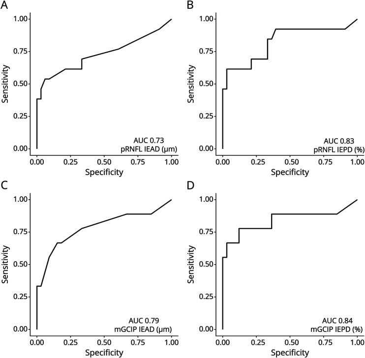
A multicenter validation study was conducted on the published IED cutoff values (>4% or >4 μm in the macular ganglion cell and inner plexiform layer [mGCIP] or >5% or >5 μm in the peripapillary retinal nerve fiber layer [pRNFL]) in individuals with MOG-ON and age-matched and sex-matched healthy controls (HCs). Structural data were acquired with Spectralis spectral-domain OCT >6 months after ON. We calculated sensitivity, specificity, and receiver operating characteristics for both intereye percentage (IEPD) and absolute difference (IEAD).

RESULTS

A total of 66 individuals were included (MOG-ON N = 33; HCs N = 33). ON was unilateral in 20 and bilateral in 13 subjects. In the pooled analysis, the mGCIP IEPD was most sensitive (92%), followed by the mGCIP IEAD (88%) and pRNFL (84%). The same pattern was found for the specificity (mGCIP IEPD 82%, IEAD 82%; pRNFL IEPD 82%, IEAD 79%).In subgroup analyses, the diagnostic sensitivity was higher in subjects with unilateral ON (>99% for all metrics) compared with bilateral ON (61%-78%).

DISCUSSION

In individuals with MOG-ON, the diagnostic accuracy of OCT-based IED metrics for ON was high, especially of mGCIP IEPD.

CLASSIFICATION OF EVIDENCE

This study provides Class III evidence that the intereye difference on OCT can distinguish between those with MOG and normal controls.

摘要

背景和目的

2022 年视神经炎国际联合会诊断标准(ON)包括光学相干断层扫描(OCT)。在多发性硬化症和水通道蛋白 4 抗体阳性视神经脊髓炎谱系障碍患者中,双眼间差异(IED)指标对 ON 的诊断价值较高,但在髓鞘少突胶质细胞糖蛋白抗体相关性 ON(MOG-ON)中尚不清楚。

方法

对已发表的 MOG-ON 患者和年龄、性别匹配的健康对照者(HCs)中 IED 截断值(黄斑神经节细胞和内丛状层[macular ganglion cell and inner plexiform layer,mGCIP]>4%或>4μm,或视盘周围视网膜神经纤维层[peripapillary retinal nerve fiber layer,pRNFL] >5%或>5μm)进行多中心验证研究。ON 后 6 个月以上采用 Spectralis 光谱域 OCT 采集结构数据。我们计算了双眼间百分比(IEPD)和绝对差值(IEAD)的敏感性、特异性和受试者工作特征。

结果

共纳入 66 例患者(MOG-ON 33 例;HCs 33 例)。20 例为单侧 ON,13 例为双侧 ON。在汇总分析中,mGCIP IEPD 最敏感(92%),其次是 mGCIP IEAD(88%)和 pRNFL(84%)。特异性也呈现相同模式(mGCIP IEPD 82%,IEAD 82%;pRNFL IEPD 82%,IEAD 79%)。在亚组分析中,单侧 ON 患者的诊断敏感性较高(所有指标均>99%),而双侧 ON 患者的诊断敏感性为 61%-78%。

讨论

在 MOG-ON 患者中,基于 OCT 的 IED 指标对 ON 的诊断准确性较高,尤其是 mGCIP IEPD。

证据分类

本研究提供 III 级证据,表明 OCT 上的双眼间差异可区分 MOG 与正常对照者。

https://cdn.ncbi.nlm.nih.gov/pmc/blobs/917f/11379125/3a3b880df758/NXI-2024-100159f4.jpg
https://cdn.ncbi.nlm.nih.gov/pmc/blobs/917f/11379125/0351ecd7361f/NXI-2024-100159f1.jpg
https://cdn.ncbi.nlm.nih.gov/pmc/blobs/917f/11379125/99465c7f9f15/NXI-2024-100159f2.jpg
https://cdn.ncbi.nlm.nih.gov/pmc/blobs/917f/11379125/eecbe536d504/NXI-2024-100159f3.jpg
https://cdn.ncbi.nlm.nih.gov/pmc/blobs/917f/11379125/3a3b880df758/NXI-2024-100159f4.jpg

相似文献

1
Diagnostic Value of Inter-Eye Difference Metrics on OCT for Myelin Oligodendrocyte Glycoprotein Antibody-Associated Optic Neuritis.OCT 上的眼间差异指标对髓鞘少突胶质细胞糖蛋白抗体相关性视神经炎的诊断价值。
Neurol Neuroimmunol Neuroinflamm. 2024 Nov;11(6):e200291. doi: 10.1212/NXI.0000000000200291. Epub 2024 Sep 4.
2
Diagnostic value of intereye difference metrics for optic neuritis in aquaporin-4 antibody seropositive neuromyelitis optica spectrum disorders.水通道蛋白 4 抗体阳性视神经脊髓炎谱系疾病中双眼差异指标对视神经炎的诊断价值。
J Neurol Neurosurg Psychiatry. 2023 Jul;94(7):560-566. doi: 10.1136/jnnp-2022-330608. Epub 2023 Feb 21.
3
Retina thickness in clinically affected and unaffected eyes in patients with aquaporin-4 immunoglobulin G antibody seropositive neuromyelitis optica spectrum disorders: a systematic review and meta-analysis.水通道蛋白4免疫球蛋白G抗体血清阳性视神经脊髓炎谱系障碍患者患眼与未患眼的视网膜厚度:一项系统评价和荟萃分析
J Neurol. 2023 Feb;270(2):759-768. doi: 10.1007/s00415-022-11482-4. Epub 2022 Nov 10.
4
Retinal layer segmentation in multiple sclerosis: a systematic review and meta-analysis.多发性硬化症中的视网膜层分割:系统评价和荟萃分析。
Lancet Neurol. 2017 Oct;16(10):797-812. doi: 10.1016/S1474-4422(17)30278-8. Epub 2017 Sep 12.
5
Optical coherence tomography and angiography reveal early retinal alterations in pediatric-onset multiple sclerosis.光学相干断层扫描和血管造影显示儿童期多发性硬化症患者早期视网膜改变。
Eur J Pediatr. 2025 Jun 6;184(7):398. doi: 10.1007/s00431-025-06234-1.
6
Ganglion Cell Layer Thickness as a Biomarker for Amyotrophic Lateral Sclerosis Functional Outcome: An OCT study.神经节细胞层厚度作为肌萎缩侧索硬化症功能预后的生物标志物:一项光学相干断层扫描研究。
Rom J Ophthalmol. 2025 Apr-Jun;69(2):200-207. doi: 10.22336/rjo.2025.32.
7
Uncommon Non-MS Demyelinating Disorders of the Central Nervous System.中枢神经系统罕见的非多发性硬化脱髓鞘疾病
Curr Neurol Neurosci Rep. 2025 Jul 1;25(1):45. doi: 10.1007/s11910-025-01432-8.
8
Optic nerve head and fibre layer imaging for diagnosing glaucoma.用于诊断青光眼的视神经乳头和纤维层成像。
Cochrane Database Syst Rev. 2015 Nov 30;2015(11):CD008803. doi: 10.1002/14651858.CD008803.pub2.
9
Optical coherence tomography (OCT) for detection of macular oedema in patients with diabetic retinopathy.光学相干断层扫描(OCT)用于检测糖尿病视网膜病变患者的黄斑水肿。
Cochrane Database Syst Rev. 2015 Jan 7;1(1):CD008081. doi: 10.1002/14651858.CD008081.pub3.
10
Optical coherence tomography angiography measurements in neuromyelitis optica spectrum disorder and myelin oligodendrocyte glycoprotein antibody disease: A systematic review and meta-analysis.视神经脊髓炎谱系疾病和髓鞘少突胶质细胞糖蛋白抗体病的光学相干断层扫描血管造影测量:系统评价和荟萃分析。
Mult Scler Relat Disord. 2024 Nov;91:105864. doi: 10.1016/j.msard.2024.105864. Epub 2024 Sep 2.

本文引用的文献

1
Prognostic factors of first-onset optic neuritis based on diagnostic criteria and antibody status: a multicentre analysis of 427 eyes.基于诊断标准和抗体状态的首发视神经炎的预后因素:427只眼的多中心分析
J Neurol Neurosurg Psychiatry. 2024 Jul 15;95(8):753-760. doi: 10.1136/jnnp-2023-333133.
2
Diagnostic value of intereye difference metrics for optic neuritis in aquaporin-4 antibody seropositive neuromyelitis optica spectrum disorders.水通道蛋白 4 抗体阳性视神经脊髓炎谱系疾病中双眼差异指标对视神经炎的诊断价值。
J Neurol Neurosurg Psychiatry. 2023 Jul;94(7):560-566. doi: 10.1136/jnnp-2022-330608. Epub 2023 Feb 21.
3
Diagnosis of myelin oligodendrocyte glycoprotein antibody-associated disease: International MOGAD Panel proposed criteria.
髓鞘少突胶质细胞糖蛋白抗体相关性疾病的诊断:国际 MOGAD 专家组提出的标准。
Lancet Neurol. 2023 Mar;22(3):268-282. doi: 10.1016/S1474-4422(22)00431-8. Epub 2023 Jan 24.
4
Details and outcomes of a large cohort of MOG-IgG associated optic neuritis.一大群与MOG-IgG相关的视神经炎的详细情况及结果
Mult Scler Relat Disord. 2022 Dec;68:104237. doi: 10.1016/j.msard.2022.104237. Epub 2022 Oct 10.
5
Diagnosis and classification of optic neuritis.视神经炎的诊断和分类。
Lancet Neurol. 2022 Dec;21(12):1120-1134. doi: 10.1016/S1474-4422(22)00200-9. Epub 2022 Sep 27.
6
Longitudinal Retinal Changes in MOGAD.MOGAD 中的纵向视网膜变化。
Ann Neurol. 2022 Sep;92(3):476-485. doi: 10.1002/ana.26440. Epub 2022 Jul 16.
7
OCT retinal nerve fiber layer thickness differentiates acute optic neuritis from MOG antibody-associated disease and Multiple Sclerosis: RNFL thickening in acute optic neuritis from MOGAD vs MS.光学相干断层扫描(OCT)测量的视网膜神经纤维层厚度可区分急性视神经炎与MOG抗体相关疾病及多发性硬化症:MOG抗体相关疾病与多发性硬化症所致急性视神经炎的视网膜神经纤维层增厚情况比较
Mult Scler Relat Disord. 2022 Feb;58:103525. doi: 10.1016/j.msard.2022.103525. Epub 2022 Jan 11.
8
Retinal inter-eye difference and atrophy progression in multiple sclerosis diagnostics.多发性硬化症诊断中的视网膜双眼差异与萎缩进展。
J Neurol Neurosurg Psychiatry. 2022 Feb;93(2):216-219. doi: 10.1136/jnnp-2021-327468. Epub 2021 Nov 11.
9
Retinal Optical Coherence Tomography in Neuromyelitis Optica.视神经脊髓炎的视网膜光学相干断层扫描。
Neurol Neuroimmunol Neuroinflamm. 2021 Sep 15;8(6). doi: 10.1212/NXI.0000000000001068. Print 2021 Nov.
10
APOSTEL 2.0 Recommendations for Reporting Quantitative Optical Coherence Tomography Studies.APOSTEL 2.0 关于报告定量光学相干断层扫描研究的建议。
Neurology. 2021 Jul 13;97(2):68-79. doi: 10.1212/WNL.0000000000012125. Epub 2021 Apr 28.