Thant May Thazin, Hasriadi Hasriadi, Poldorn Preeyaporn, Jungsuttiwong Siriporn, Rojsitthisak Pornchai, Böttcher Chotima, Towiwat Pasarapa, Sritularak Boonchoo
Department of Pharmacognosy and Pharmaceutical Botany, Faculty of Pharmaceutical Sciences, Chulalongkorn University Bangkok 10330 Thailand
Department of Pharmacology and Physiology, Faculty of Pharmaceutical Sciences, Chulalongkorn University Bangkok 10330 Thailand.
RSC Adv. 2024 Sep 5;14(39):28390-28400. doi: 10.1039/d4ra04761c. eCollection 2024 Sep 4.
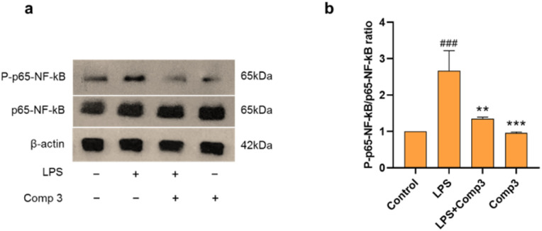
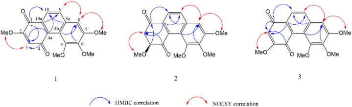
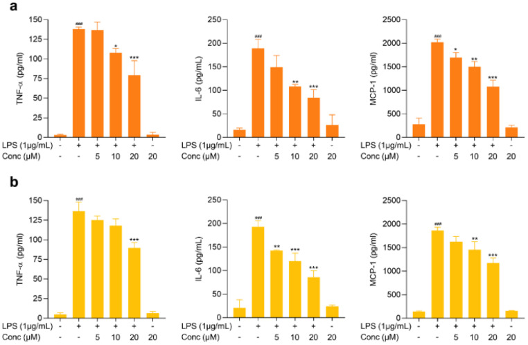
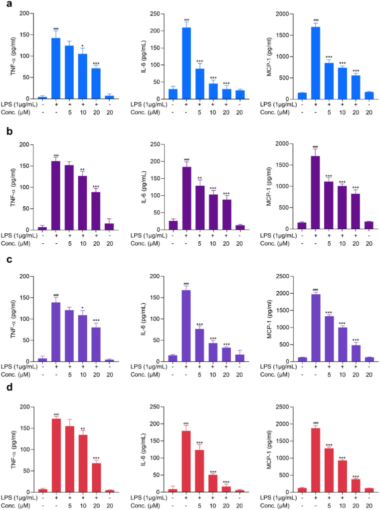
The roots of yielded a total of 17 compounds, comprising two new compounds (1-2), one new natural product (3), and 14 known compounds (4-17). The structures of new compounds were determined through the analysis of their spectroscopic data, including NMR, MS, UV, FT-IR, optical rotation, and CD. The anti-inflammatory activity of the isolated pure compounds was assessed using lipopolysaccharide-activated BV2 microglial cells. Compounds 1, 3, 6, 12, 14, and 16 showed the ability to reduce LPS induced NO release in BV2 microglial cells, with IC values of 9.95 ± 2.13, 8.77 ± 3.78, 2.39 ± 0.91, 6.69 ± 2.94, 2.96 ± 1.38, 8.42 ± 2.99 μM, respectively and reduced the secretion of proinflammatory mediators (TNF-α, IL-6, MCP-1) in a concentration-dependent manner. Furthermore, the mechanistic role of the compound 3 was determined, which demonstrated its ability to inhibit the nuclear factor-κB (NF-κB) pathway through decreasing phosphorylation of p65 subunits.
其根中共产生了17种化合物,包括两种新化合物(1 - 2)、一种新天然产物(3)和14种已知化合物(4 - 17)。通过对其光谱数据的分析确定了新化合物的结构,这些光谱数据包括核磁共振(NMR)、质谱(MS)、紫外光谱(UV)、傅里叶变换红外光谱(FT - IR)、旋光度和圆二色光谱(CD)。使用脂多糖激活的BV2小胶质细胞评估分离出的纯化合物的抗炎活性。化合物1、3、6、12、14和16显示出能够降低脂多糖诱导的BV2小胶质细胞中一氧化氮(NO)的释放,其半数抑制浓度(IC)值分别为9.95±2.13、8.77±3.78、2.39±0.91、6.69±2.94、2.96±1.38、8.42±2.99微摩尔,并以浓度依赖的方式减少促炎介质(肿瘤坏死因子 -α、白细胞介素 - 6、单核细胞趋化蛋白 - 1)的分泌。此外,还确定了化合物3的作用机制,它通过降低p65亚基的磷酸化来证明其抑制核因子 -κB(NF -κB)途径的能力。